A. Development Of The Hoof. Part 2
Description
This section is from the "Diseases Of The Horse's Foot" book, by H. Caulton Reeks. Also available from Amazon: Diseases Of The Horse's Foot.
A. Development Of The Hoof. Part 2
The hair grows from the bottom of the follicle by a multiplication of the cells covering the papilla upon which its root is moulded. When a hair is cast off a new one is produced from the cells covering the papilla, or, in case of the death or degeneration of the original papilla, the new hair is produced from a second papilla formed in place of the first at the bottom of the follicle.
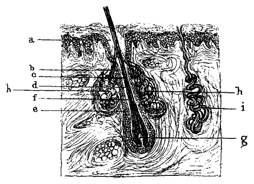
Fig. 24. - Section Of Skin With Hair Follicle And Hair. A, The Hair Follicle; B, The Hair Root; C, The Medulla; D, The Hair Cuticle; E, The Outer Root Sheath; F, The Inner Root Sheath; G, The Papilla From Which The Hair Is Growing; H, A Sebaceous Gland; I, A Sudoriferous Gland.
The Sebaceous Glands are small saccular glands with their ducts opening into the mouths of the hair follicles. They furnish a natural lubricant to the hairs and the skin.
The Sudoriferous Or Sweat Glands are composed of coiled tubes which lie in the deeper portion of the skin, and send up a corkscrew-like duct to open on the surface of the epidermis. They are numerous over the whole of the body.

Fig. 25. - Longitudinal Section Through Nail And Nail-Bed Of A Human Foetal Finger.[A] A, The Nail; B, The Rete Mucosum; C, The Longitudinal Ridges Of The Corium.
[Footnote A: Seeing that the section is a longitudinal one, it would appear from the way the ridges cut that they are running transversely beneath the nail. Their extreme delicacy, however, prevents a single one showing itself along the length of the section, and their constant accidental cutting makes them appear to run transversely (H.C.R.).]
The Human Nails are thickenings of the lowermost layer of the horny portion of the epidermis, the stratum lucidum. They are developed over a modified portion of the corium known as the nail-bed. The horny substance of the nail is composed of clear horny cells, and rests immediately upon a Malpighian layer similar to that found in the epidermis generally. Instead of the papillae present elsewhere in the skin, the corium of the nail-bed is marked by longitudinal ridges, a similar, though less distinct, arrangement to that found in the laminae of the horse's foot.
Having thus paved the way, we are now in a better position to discuss our original question (Are the horny laminae secreted by the sensitive?), and better able to appreciate the work that has been done towards the elucidation of the problem.
A most valuable contribution to this study is an article published in 1896 by Professor Mettam.[A] Here the question is dealt with in a manner that must effectually silence all other views save such as are based upon similar methods of investigation - namely, histological examination of sections of equine hoofs in various stages of foetal development.
[Footnote A: The Veterinarian, vol. lxix., p.1.]
Professor Mettam commences by drawing attention to the error that has been made in this connection by studying the soft structures of the foot separated by ordinary putrefactive changes from the horny covering. "In this way," the writer points out, "a wholly erroneous idea has crept in as to the relation of the one to the other, and the two parts have been treated as two anatomical items, when, indeed, they are portions of one and the same thing. As an illustration, and one very much to the point at issue, the soft structures of the foot are to the horny covering what the corium of the skin and the rete Malpighii are to the superficial portions of the epidermis. Indeed, the point where solution of continuity occurs in macerating is along the line of the soft protoplasmic cells of the rete."
In the foregoing description of the skin we have seen that the corium is not a plane surface, but that it is studded by numerous papillary projections, and that these projections, with the depressions between them, are covered by the cells of the epidermis.
The corium of the horse's foot, however, although possessed of papillae in certain positions (as, for example, the papillae of the coronary cushion, and those of the sensitive frog and sole), has also most pronounced ridges (laminae) which run down the whole depth of the os pedis. Each lamina again carries ridges (laminellae) on its lateral aspects, giving a section of a lamina the appearance of being studded with papillae. We have already pointed out the ridge-like formation of the human nail-bed, and noted that, with the exception that the secondary ridges are not so pronounced, it is an exact prototype of the laminal formation of the corium of the horse's foot.
The distribution of the laminae over the foot we have discussed in the chapter devoted to the grosser anatomy. In a macerated foot the sensitive laminae of the corium interdigitate with the horny laminae of the hoof; that is to say, there is no union between the two, for the simple reason that it has been destroyed; they simply interlock like the unglued junction of a finely dovetailed piece of joinery. But no further, however, than the irregularities of the underneath surface of the epidermis of the skin can be said to interlock with the papillae of the corium does interlocking of the horny and sensitive laminae occur. It is only apparent. The horny laminae are simply beautifully regular epidermal ingrowths cutting up the corium into minute leaf-like projections.
In a macerated specimen, then, the exposed sensitive structures of the foot exhibit the corium as (1) the Coronary Cushion, fitting into the cutigeral groove; (2) the Sensitive Laminae, clothing the outer surface of the terminal phalanx, and extending to the bars; (3) the Plantar Cushion, or sensitive frog; and (4) the Sensitive Sole.
Continue to:
My Books

