Uterus (fig. 230)
Description
This section is from the book "The Horse - Its Treatment In Health And Disease", by J. Wortley Axe. Also available from Amazon: The Horse. Its Treatment In Health And Disease.
Uterus (fig. 230)
This is the organ which receives the ovum and in which the foetus is developed. It is composed of a neck and a body, which divides into two horns of approximately equal size.
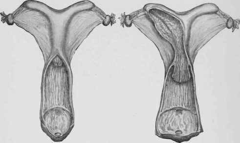
Fig. 230. - Uteri with Short and Long Necks.
The neck is embraced by the anterior extremity of the vagina, into which it projects after the manner of the end of a tap into a barrel; it is rounded in outline though somewhat compressed from above downwards. The opening of the neck into the vagina is called the " os uteri externum". The opposite extremity opens into the body of the organ, and is the "os uteri internum". The body of the uterus is about 8 to 10 inches in length, and is situated partly within the pelvis and partly in the abdomen, having the rectum above and the bladder below. The horns are about 8 inches in length, and hang suspended by two broad ligaments from the lumbar portion of the spine. They are somewhat laterally compressed, are concave on their upper surface and convex on the lower. The anterior extremity is obtusely pointed, and receives the Fallopian tubes. The opposite extremity is continuous with the body of the organ.
Structure
The wall of the uterus is composed of the following structures arranged in the following order from without inwards: - (1) serous or peritoneal covering, which invests the entire organ with the exception of the cervix or neck. (2) Muscular coat, composed of longitudinal and circular fibres. (3) Sub-mucous areolar tissue. (4) Mucous membrane raised into longitudinal folds and containing numerous tubular glands.
Attachments
The whole uterus, with the exception of that portion of the neck within the vagina, is covered with peritoneum, which, branching off from the organ in various directions, forms the ligaments by which it is attached to the rectum above, the bladder below, and to the sides of the pelvis and the roof of the belly.
The recto-vaginal ligament leaves the lower face of the rectum and passes on to the upper surface of the vagina and uterus. The vesicovaginal ligament leaves the lower surface of the uterus and vagina and passes on to the upper face of the bladder. The broad ligaments are simply the lateral edges of the folds reflected from the sides of the organ and attached at first to the sides of the pelvis, from which they rise to the roof of the abdomen and support the uterine horns. Hence it will be seen that the lateral ligaments form a horizontal partition (fig. 229, e) composed of an upper and a lower layer between which the uterus is placed. The only other ligament of any importance is the round ligament (fig. 231, g). This is a fibro-muscular cord situated between the layers of the broad ligament, and is attached to the concavity of the upper part of the horn of the uterus and to the floor of the abdomen in front of the pubic bone. The uterus, therefore, is supported and held in position (l) by the projection of its neck into the vagina, (2) by a peritoneal fold connecting it to the rectum above, (3) to the bladder below, and (4) to the sides of the pelvis.
Between the layers of the broad ligaments there are several other structures of importance in relation to the generative function, about some of which something must be said. There are (1) the Fallopian tubes, (2) ovarian ligaments, (3) the ovaries and the vessels and nerves distributed thereto and to the uterus.
The Fallopian tube (fig. 231, b) is contained in the anterior margin of the broad ligament. It extends from the ovary to the extremity of the horn of the uterus. It is 8 or 9 inches long, very tortuous, and through it the eggs are conducted from the ovary or egg-forming gland to the uterus. The uterine extremity of the tube opens into the uterus by a minute aperture scarcely larger than an ordinary pinhole. The other end opens into the abdominal cavity, and is attached by about 1/2 inch of its circumference to the ovary (fig. 231). It is trumpet-shaped and surrounded by a fringe of mucous membrane, from which it has been called the " fimbriated extremity ".
The Ovary (fig. 231, f) is oval in form and laterally compressed, pale pink in colour, and weighs about 3 ounces. It hangs in the abdominal cavity, suspended in a special pouch from the internal layer of the broad ligament; its surface is usually very uneven, from the bulging of small cysts, and is scarred here and there, marking the points at which other cysts have come and gone. It has two curvatures; the greater or convex border is turned upwards and somewhat backwards, and the lesser curvature or "hilum" looks downward and a little forward. This is the attached part and receives the blood-vessels, etc. The two extremities or poles are anterior and posterior; the former gives attachment to the Fallopian tube while the latter is connected with the ovarian ligament. This ligament, composed of fibrous tissue and unstriated muscle, extends from the free extremity of the horn of the uterus to the ovary, and occupies the internal of the two layers which form the anterior margin of the broad ligament.
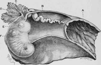
Fig. 231. - Fallopian Tube connecting the Uterus with the Ovary.
A, Uterus. B, Fallopian Tube. c, Its Fimbriated extremity. D. Opening into the Tube, through which the Ovum or Egg E, passes from the Ovary F. G, Round Ligament of the Ovary.
The essential portion of the gland consists of small bladder-like bodies termed "Graafian follicles" or ovisacs (fig. 232, E, g). These, when fully developed, are filled with fluid and contain the eggs. A young Graafian follicle consists of a spherical cell, the ovum, closely invested by a single layer of epithelium; these may be seen forming an almost continuous layer near the surface of the ovary. In the deeper parts of the gland, that is to say nearer the hilum, the more developed and larger follicles will be noticed.
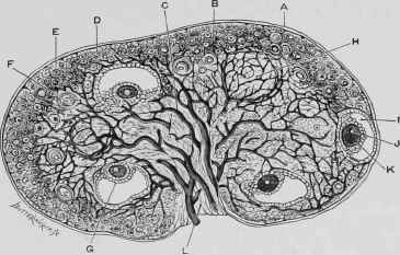
Fig. 232. - The Ovary.
A, Peritoneal Coat. B, D, E, F, Cortical Vesicles, c, Blood-vessels. G, Graafian Follicle. H, Stroma. I, Graafian Follicle. J, Germinal Spot. K, Germinal Vesicle. L, Attached border.
The fully developed or ripe follicles (size about 1/8 inch) are surrounded by a fibrous wall, the "tunica fibrosa", containing a capillary plexus. This is lined by several layers of epithelium, " tunica granulosa", which in one part is elevated into a little mound, "discus proligerus", in which the ovum is embedded; the rest of the cavity, by far the greater part, is filled with an albuminous fluid, " liquor folli-culi". Pathological cysts are frequently seen in the ovaries, especially of cows and mares, varying in size from small marbles to oranges. These arise in the corpora lutea, and are not to be mistaken for the Graafian follicles.
Continue to:
My Books

