Annelida. Part 23
Description
This section is from the book "General Outline Of The Organization Of The Animal Kingdom, And Manual Of Comparative Anatomy", by Thomas Rymer Jones. Also available from Amazon: A General Outline of the Animal Kingdom and Manual of Comparative Anatomy.
Annelida. Part 23
(689). At the point corresponding with the circular vessel (fig. 134, 5), the primary ventral sends off a considerable division for the supply of the intestinal system. The current, therefore, entering the glandular parietes of the intestine is purely arterial in this genus; for it is unmixedly composed of blood returning from the tentacles and branchiae, by both of which the function of respiration is performed. Here, again, there exist but two principal directions in which the blood circulates, viz. longitudinally and transversely, or circularly, the former currents being connected with the latter. The circular vessel (fig. 134, b) acts like an auricle; it receives the blood from the intestinal system and delivers it into the great dorsal (a.) The alimentary canal is embraced in this genus, as in all Annelids, by a framework of longitudinal and transverse vessels (f), in which the blood moves backwards below, and forwards above.
(690). In the dissection of Terebella nebu-losa, figured by Milne-Edwards, a large vessel (fig. 135,1) is readily distinguishable towards the anterior part of the animal, running along the median line of the back, and situated immediately beneath the integuments. This vessel rests upon the alimentary canal, and exhibits irregular contractile movements whereby the blood contained in its interior is propelled from behind forwards, and consequently performs the functions of a heart; and if we would compare it with what exists in the higher animals, it might be considered as physiologically representing a pulmonic ventricle, seeing that the vessels that convey the blood to the branchiae for the purposes of respiration take their origin from its anterior extremity.
(691). By its posterior extremity, the great dorsal trunk receives the blood which it is appointed to propel through the branchial organs, from several large veins which are, for the most part, adherent to the walls of the intestine (fig. 134,f), from which they receive a multitude of branches derived from the rich vascular network distributed over the intestinal walls. The principal veins, however, that communicate with this tubular heart are two large transverse trunks which form a ring around the digestive canal, beneath which they unite and become continuous with a large median trunk (fig. 135, h) that runs along the under surface of the intestine, from which, in the same manner as the dorsal veins, it receives numerous lateral branches derived from the vascular network already mentioned. Lastly, there is a small median trunk, situated upon the internal surface of the integuments of the back (fig. 135, m), into which open the veins derived from the different segments of the body, and which likewise communicates with the dorso-intestinal vessel by numerous anastomosing ramifications.
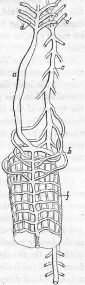
Fig. 134. Respiratory and circulatory apparatus in Terebella. (After Dr. Williams).
(692). The vessels above enumerated may be considered as constituting the general venous system of the body; and the blood which they convey to the dorsal trunk is, by the contractions of that vessel, in great part distributed to the branchiae through three pairs of branchial arteries derived immediately from the dorsal heart. Still, however, all the blood thus moving from behind forwards is not conveyed to the branchial organs, since a certain portion finds its way through a small median vessel to the labial organs and cephalic ten-tacula.
(693). After having passed through the branchial organs, the renovated blood is received by vessels which unite to form a median trunk (fig. 135, o) that runs beneath the alimentary tube and immediately above the ventral chain of nervous ganglia. This ventral trunk is continued along the whole length of the body, and gives off opposite to each ring a pair of transverse vessels, which, after having supplied branches to the integument and locomotive organs, bend upwards, to be distributed over the walls of the intestine, where their ramifications contribute to form the vascular network above alluded to.
(694). The ventral vessel and its ramifications fulfil, therefore, the functions of an arterial system; and consequently the branchiae themselves must be regarded as the agents employed in propelling the blood through the systemic circulation. These organs, indeed, may be observed, at intervals, to contract with considerable energy, and thus materially to assist in urging the blood through the arterial ramifications.
(695). M. de Quatrefages observes* that both in the Erratic andTubi-colous Annelidans the sexes are separate, and states that the generative apparatus, both in the males and females, is restricted to the abdominal portion of the body. According to this distinguished anatomist, the testicle consists of a kind of areolar web of extreme delicacy, which, arising from a median aponeurosis, adheres to the internal and inferior surface of the general cavity, rising as high as the middle of the digestive canal. The tenuity of this tissue is such that it is impossible to procure more than small fragments for microscopic examination.
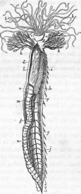
Fig. 135. Arrangement of the vascular system in Terebella. (After Milne-Edwards).
* "Memoire sur les Hermelliens," Ann. des Sci. Nat. 3 ser. 1848.
(696). The ovary is in every respect similar to the testicle: perhaps its texture may be rather firmer, but not sufficiently so to be adapted for satisfactory histological distinction.
(697). In the males as well as in the females, but more especially in the latter, during the period of reproduction a pigment is secreted in great abundance, which lines the generative organs; but in proportion as the ova or zoosperms become developed, the amount of this pigment diminishes. Both the ovary and testicle are evidently temporary organs, no traces of them being distinguishable in the generality of specimens; and moreover, in proportion as their products become developed in the general visceral cavity, they become gradually atrophied. When the male secretion is at maturity, a jet of water washes away the sperma-tozoids, and no trace of the testicle is left; on the contrary, when the sperm is immature, washing still leaves behind a delicate web almost resembling a light cloud.
Continue to:
My Books

