Chapter XVI. Cirrhopoda
Description
This section is from the book "General Outline Of The Organization Of The Animal Kingdom, And Manual Of Comparative Anatomy", by Thomas Rymer Jones. Also available from Amazon: A General Outline of the Animal Kingdom and Manual of Comparative Anatomy.
Chapter XVI. Cirrhopoda
(1164). The Cirrhopoda present a strange combination of articulated limbs united with many of the external characters of a Mollusk, as will be at once evident from the examination of any species of Barnacle, whether sessile or pedunculated. We select a common form, Pentelasmis vitrea, as an example of the kind last mentioned. The animal in question is enclosed in a shell resembling in some respects that of the common Mussel, but composed of five distinct pieces united together by a dense intervening membrane: of these, four pieces are lateral, and disposed in pairs; while a fifth, which is single, is interposed between the posterior edges of the two valves, so as to unite them along the whole length of the back. Along the anterior margin the valves are only partially connected by membrane; so that a long fissure is left, through which the articulated extremities may be protruded. In place of the hinge that joins the two shells of the Mussel, we find the tough coriaceous membrane that unites the different shelly pieces of the integument of Pentelasmis prolonged into a cylindrical pedicle (fig. 235, I), which is in some species many inches in length, and, being attached by its extremity to any submarine body, fixes the animal permanently to the same locality. The external layer of this pedicle is coriaceous, or almost corneous, in its appearance, being evidently an epidermic structure; but internally the tube is lined with a layer of strong muscular fibres arranged longitudinally (fig. 235, m, ri), which by their contraction are no doubt able to bend the flexible stem in any given direction, and thus confer upon the animal a limited power of changing its position when necessary. On removing one-half of the shelly covering (as in fig. 232, a a), we expose the body of the Cirrhopod, and discern the following particulars. The lower portion of the body, which encloses the principal viscera (b b), is soft and much dilated, especially towards the dorsal region; this part of the animal is covered with a delicate membrane, beneath which is a layer of whitish granular substance. The mouth (g) is seen upon the ventral aspect, situated immediately at the inferior extremity of that longitudinal fissure in the mantle through which the arms are protruded: the oral aperture appears to be raised upon a prominent tubercle, and, when attentively examined, is found to be provided with a rudimentary apparatus of jaws, presenting a distinct lip, furnished with minute palpi, and three pairs of mandibles, of which the two external are horny and serrated, while the third remains permanently soft and membranous. Immediately behind the mouth, we find on each side certain pyramidal fleshy appendages (d d d), resembling, as Hunter expressed it, a minute Starfish, which no doubt constitute the branchial or respiratory organs.
Commencing above the mouth, we further notice on each side six pairs of articulated and flexible arms or cirri (fig. 232, c c), each being composed of a series of semi - corneous pieces, and exhibiting at each joint long and stiff hairs. Every pair of cirri arises from a single prominent stem; and those most distant from the mouth being the longest and most extensile, the whole apparatus, consisting of twenty-four cirri, forms, when protruded from the body, a kind of net of exquisite contrivance, in which passing particles of nourishment are easily entangled, and thus conveyed to the mouth. Lastly, on separating the cirriferous pedicles, we find, terminating the body, and forming, as it were, a kind of tail, a long, soft, and flexible organ (fig. 235, h), the extremity of which is perforated by a minute aperture; but the real nature of this instrument we shall examine by and by.
(1165). On reviewing this general description of the external construction of Pentelasmis, the reader cannot but be struck with the singular combination of characters which it exhibits. Judging from its shell alone, its right to be considered a Mollusk would seem to be at once demonstrable; for, in fact, most conchologists agree in claiming these animals as belonging to their own department; and yet if, after removing the shell, we compare the animal with a Crustacean, its alliance with that class is equally evident. Suppose the body (fig. 232, b b) to represent the thoracic portion of a Crustacean slightly bent upon itself, and enclosed in an extensively developed thorax*; the valves of the shell would represent this thorax, which would be divided into five pieces; the first pair of cirri arising from the body would then represent the true feet of a Crustacean; the branchiae would occupy the same position in both; the rest of the body of the Barnacle, namely that which supports the five other pairs of feet, would represent the tail of the Crustacean, and the ciliated, natatory feet, generally connected with that part of the external skeleton. Even the mouth, as the author referred to might have added, with its triple series of jaws, is more nearly allied in structure to that of the Crustaceans than to anything we shall meet with in the structure of the oral organs of true Mollusca.
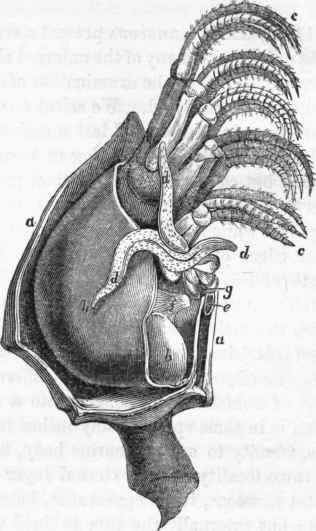
Fig. 232. Pentelasfnia vitrea: a a, the shelly valves; b b, body contained within the shells; c c, the cirri; d d d, presumed branchial apparatus; e,f, muscular expansions; g, the mouth. (After Hunter).
* Cuvier, Memoire sur les Animaux des Anatifes et des Balanes, et sur leur Anatomie, p. 6.
(1166). But the affinity which unites the Cirrhopoda to the Homo-gangliata is not merely exemplified in the analogies that can be pointed out between the external configuration of Pentelasmis and some Crustacean forms; the nervous system even, as we might be led to anticipate from the symmetrical arrangement of the articulated cirri, still exhibits the Homo-gangliate condition, and, besides the supra-cesophageal masses, forms a longitudinal chain of double ganglia arranged along the ventral surface of the body, from which the nerves supplying the cirriferous arms take their origins. Four small tubercles (fig. 233)*, placed transversely above the oesophagus, represent the brain, and give origin to four principal nerves (ffff), which are distributed to the muscles and viscera; for in such a situation, organs of sense would evidently be useless. Two lateral cords, derived from the above, surround the oesophagus, from each of which a nerve (o o) is given off. Below the oesophagus the nervous collar terminates in a pair of ganglia (h), that give origin to the nerves supplied to the first pair of arms; and then succeeds a parallel series of double ganglia (i, k, I, m), exactly resembling those of articulated animals, from which emanate nerves that are destined to the cirri and surrounding parts.
Continue to:
My Books

