The Life History Of Vaucheria
Description
This section is from "Scientific American Supplement". Also available from Amazon: Scientific American Reference Book.
The Life History Of Vaucheria
By A.H. BRECKENFELD.
Nearly a century ago, Vaucher, the celebrated Genevan botanist, described a fresh water filamentous alga which he named Ectosperma geminata, with a correctness that appears truly remarkable when the imperfect means of observation at his command are taken into consideration. His pupil, De Candolle, who afterward became so eminent a worker in the same field, when preparing his "Flora of France," in 1805, proposed the name of Vaucheria for the genus, in commemoration of the meritorious work of its first investigator. On March 12, 1826, Unger made the first recorded observation of the formation and liberation of the terminal or non-sexual spores of this plant. Hassall, the able English botanist, made it the subject of extended study while preparing his fine work entitled "A History of the British Fresh Water Algae," published in 1845. He has given us a very graphic description of the phenomenon first observed by Unger. In 1856 Pringsheim described the true sexual propagation by oospores, with such minuteness and accuracy that our knowledge of the plant can scarcely be said to have essentially increased since that time.
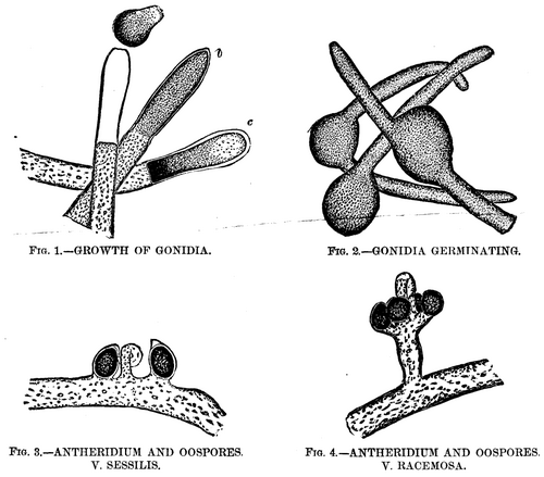
GROWTH OF THE ALGA, VAUCHERIA, UNDER THE MICROSCOPE.
Vaucheria has two or three rather doubtful marine species assigned to it by Harvey, but the fresh water forms are by far the more numerous, and it is to some of these I would call your attention for a few moments this evening. The plant grows in densely interwoven tufts, these being of a vivid green color, while the plant is in the actively vegetative condition, changing to a duller tint as it advances to maturity. Its habitat (with the exceptions above noted) is in freshwater - usually in ditches or slowly running streams. I have found it at pretty much all seasons of the year, in the stretch of boggy ground in the Presidio, bordering the road to Fort Point. The filaments attain a length of several inches when fully developed, and are of an average diameter of 1/250 (0.004) inch. They branch but sparingly, or not at all, and are characterized by consisting of a single long tube or cell, not divided by septa, as in the case of the great majority of the filamentous algae. These tubular filaments are composed of a nearly transparent cellulose wall, including an inner layer thickly studded with bright green granules of chlorophyl.
This inner layer is ordinarily not noticeable, but it retracts from the outer envelope when subjected to the action of certain reagents, or when immersed in a fluid differing in density from water, and it then becomes distinctly visible, as may be seen in the engraving (Fig. 1). The plant grows rapidly and is endowed with much vitality, for it resists changes of temperature to a remarkable degree. Vaucheria affords a choice hunting ground to the microscopist, for its tangled masses are the home of numberless infusoria, rotifers, and the minuter crustacea, while the filaments more advanced in age are usually thickly incrusted with diatoms. Here, too, is a favorite haunt of the beautiful zoophytes, Hydra vividis and H. vulgaris, whose delicate tentacles may be seen gracefully waving in nearly every gathering.
Reproduction In Vaucheria
After the plant has attained a certain stage in its growth, if it be attentively watched, a marked change will be observed near the ends of the filaments. The chlorophyl appears to assume a darker hue, and the granules become more densely crowded. This appearance increases until the extremity of the tube appears almost swollen. Soon the densely congregated granules at the extreme end will be seen to separate from the endochrome of the filament, a clear space sometimes, but not always, marking the point of division. Here a septum or membrane appears, thus forming a cell whose length is about three or four times its width, and whose walls completely inclose the dark green mass of crowded granules (Fig. 1, b). These contents are now gradually forming themselves into the spore or "gonidium," as Carpenter calls it, in distinction from the true sexual spores, which he terms "oospores." At the extreme end of the filament (which is obtusely conical in shape) the chlorophyl grains retract from the old cellulose wall, leaving a very evident clear space. In a less noticeable degree, this is also the case in the other parts of the circumference of the cell, and, apparently, the granular contents have secreted a separate envelope entirely distinct from the parent filament.
The grand climax is now rapidly approaching. The contents of the cell near its base are now so densely clustered as to appear nearly black (Fig. 1, c), while the upper half is of a much lighter hue and the separate granules are there easily distinguished, and, if very closely watched, show an almost imperceptible motion. The old cellulose wall shows signs of great tension, its conical extremity rounding out under the slowly increasing pressure from within. Suddenly it gives way at the apex. At the same instant, the inclosed gonidium (for it is now seen to be fully formed) acquires a rotary motion, at first slow, but gradually increasing until it has gained considerable velocity. Its upper portion is slowly twisted through the opening in the apex of the parent wall, the granular contents of the lower end flowing into the extruded portion in a manner reminding one of the flow of protoplasm in a living amoeba. The old cell wall seems to offer considerable resistance to the escape of the gonidium, for the latter, which displays remarkable elasticity, is pinched nearly in two while forcing its way through, assuming an hour glass shape when about half out.
The rapid rotation of the spore continues during the process of emerging, and after about a minute it has fully freed itself (Fig 1, a). It immediately assumes the form of an ellipse or oval, and darts off with great speed, revolving on its major axis as it does so. Its contents are nearly all massed in the posterior half, the comparatively clear portion invariably pointing in advance. When it meets an obstacle, it partially flattens itself against it, then turns aside and spins off in a new direction. This erratic motion is continued for usually seven or eight minutes. The longest duration I have yet observed was a little over nine and one-half minutes. Hassall records a case where it continued for nineteen minutes. The time, however, varies greatly, as in some cases the motion ceases almost as soon as the spore is liberated, while in open water, unretarded by the cover glass or other obstacles, its movements have been seen to continue for over two hours.
Continue to:
My Books

