The Abdominal Viscera
Description
This section is from the book "Applied Anatomy: The Construction Of The Human Body", by Gwilym G. Davis. Also available from Amazon: Applied anatomy: The construction of the human body.
The Abdominal Viscera
The abdominal contents should first be studied as to their positions and general relations, so that they can be readily found and identified, and then studied as to their intimate relations to the immediate surrounding structures.
By knowing the first, an operator is enabled to expose quickly the affected part, and by knowing the second he is enabled to carry out the desired procedures. While it is true that the presence of tumors or enlargement of the various organs may distort and displace them and so render their exposure and recognition difficult, nevertheless a knowledge of the normal relations is essential in order to solve the difficulties which arise in operating for or studying the various abdominal diseases and injuries.
It must be borne in mind that the extent and position of the various organs is not always the same, even though they are not diseased; it is easier to find a distended than a contracted stomach; in some people the liver though not diseased may be lower than in others, etc.
When the abdominal cavity is freely opened the general relation of the organs is visible as in Fig. 414. In the upper portion is seen the liver. Its edge usually is inclined upward toward the left, but sometimes it passes almost transversely across. In the male its lower edge should be about even with the lower edge of the thorax (tenth rib) but in females it may be a finger-breadth lower. Its anterior edge is marked by the gall-badder and round ligament. The gall-bladder is liable to be a little to the outside of its normal position at the upper extremity of the right linea semilunaris. The round ligament reaches the liver not at the median line but 2.5 to 4 cm. (1 to 1 1/2 in.) to its right. The point at which the liver crosses the median line is approximately 4 cm. (1 1/2 in.) below the tip of the ensiform cartilage. The stomach is seen to the left of the liver, between it and the left costal cartilages. Frequently the stomach is seen to pass a little to the right of the median line, particularly if it is distended. A small portion only, 2.5 to 4 cm. (1 to 1 1/2 in.), is seen in the median line and its lower border slopes up and to the left to disappear under the edge of the ribs. Immediately below the stomach lies the transverse colon, concealed beneath omentum. The omentum hangs down from the lower edge of the stomach and spreads over almost the whole of the abdomen below. It is almost always encountered in operating for appendicitis and is often found in a hernia. The gallbladder is almost the only organ below the liver and stomach which it is not liable to cover. Not infrequently the omentum is not found spread out, but from the movements of the intestines it may lie between their coils or be displaced largely to the left. The transverse colon passes upward and to the left; it crosses the median line just below the stomach and may reach as low as the umbilicus. Not infrequently, however, there may be a coil of small intestine between the level of the umbilicus and the transverse colon, or a coil may even push the transverse mesocolon in front of it and show itself between the stomach above and the transverse colon below.
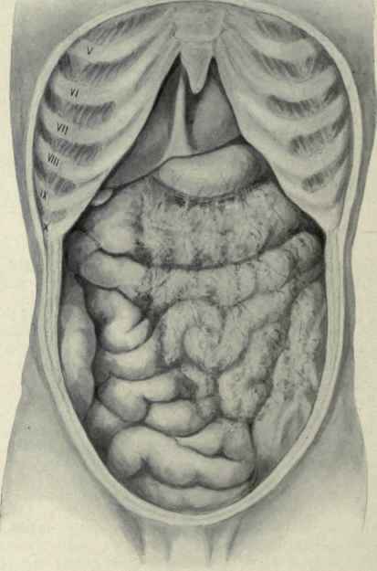
Fig. 414. - View of the abdominal organs in situ. Beneath the ensiform process is seen the liver with the round ligament to the right of the median line, below come the stomach, then the transverse colon, and lower down the small intestines, over which is spread the great omentum. In the right iliac region is seen the ascending colon and in the left the termination of the descending colon.
The caecum and the commencement of the ascending colon are almost always seen superficially in the right iliac fossa. The lower end of the caecum may reach as far forward as the middle of the inguinal (Poupart's) ligament, but when the ascending colon reaches the upper edge of the iliac crest it sinks backward out of sight, to reappear again above at the commencement of the transverse colon just below the gall-bladder.
The descending colon and sigmoid flexure are usually seen lying close to the abdominal wall somewhere between the left iliac crest and approximately the middle of Poupart's ligament. The amount visible is variable, - sometimes a considerable length is seen, at others only a single knuckle. Their presence and location are more uncertain than are those of the caecum and ascending colon on the right side. The small intestines fill the rest of the visible space. They enter the pelvis, usually are found in hernial sacs, and cover both the ascending and descending colon in the flanks. The coils in the upper and left portions of the abdomen are more likely to be jejunum, those in the lower and right portions are more likely to be ileum. Either may be found in the pelvis.
Continue to:
My Books

