Tibia
Description
This section is from the book "Handbook Of Anatomy For Students Of Massage", by Margaret E. Bjorkegren. Also available from Amazon: Handbook Of Anatomy For Students Of Massage.
Tibia
The Tibia is the inner bone of the leg. It is a long bone with a shaft and two extremities, and articulates above with the condyles of the femur and the upper end of the fibula ; below, with the lower end of the fibula and one tarsal bone, the astragalus.
The Upper Extremity is much expanded, and forms two tuberosities to support the condyles of the femur. The outer one somewhat overhangs the shaft. On the upper aspects of these are two smooth semicircular surfaces for the articular menisci, and between them in the centre a rough elevation, the intercondyloid spine. Where the outer tuberosity over-hangs the shaft is a small articular facet for the head of the fibula. On the posterior surface of the inner tuberosity is a short horizontal groove for the semimembranosus muscle. In front of the lower part of the two tuberosities is a large tubercle, the upper half of which is smooth and covered by a bursa; the lower, rough, for the attachment of the ligamentum patellae.
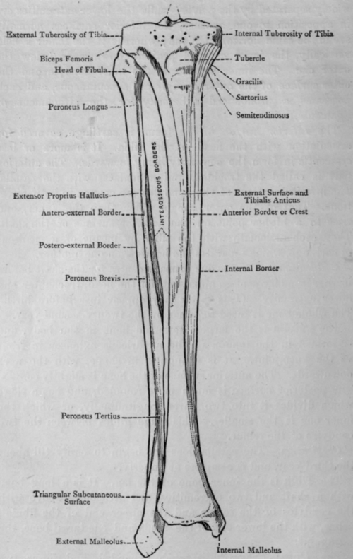
Fig. 14. - Tibia and Fibula (Anterior Surfaces).
The Shaft is triangular in shape, having an anterior, internal, and external borders, and internal, external, and posterior surfaces. The anterior border is sharp and subcutaneous, forming what is known as the shin. The internal surface is also subcutaneous, except in its upper fourth. On the posterior surface is seen the oblique, or popliteal, line, which runs from the outer tuberosity to the internal border at the junction of the upper and middle thirds. The posterior surface is divided into two parts by a vertical line dropped from the middle of the oblique line.
The Lower Extremity is expanded and becomes quadrilateral. The external surface has a large articular area for the lower end of the fibula, and the internal surface is continued downwards into a triangular process, called the internal malleolus, whose outer surface is confluent with the inferior surface of the shaft, and articulates with the astragalus. On the posterior surface just external to the malleolus there are two grooves for the passage of tendons.
Ossification
At birth the shaft is almost completely ossified, and a centre has appeared in the upper extremity. Very early the centre appears for the lower extremity, and the whole bone is fused together by the twenty-fourth year.
Continue to:
My Books

