Hydrozoa. Part 4
Description
This section is from the book "General Outline Of The Organization Of The Animal Kingdom, And Manual Of Comparative Anatomy", by Thomas Rymer Jones. Also available from Amazon: A General Outline of the Animal Kingdom and Manual of Comparative Anatomy.
Hydrozoa. Part 4
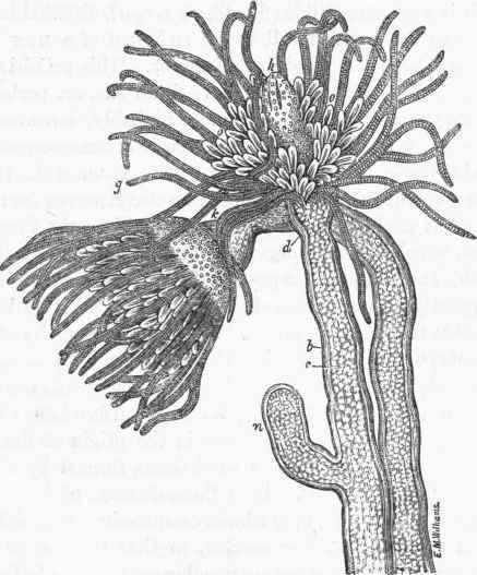
Fig. 42. Tubularia coronata. b, the polypary, or horny sheath; c, living substance of the animal; d, boundary between the individual and the common stock; g, tentacular arms; k, tentacular zone; h, mouth; n, reproduction by continuous gemmation; o, ovigerous capsules. (After Van Beneden).
(200). Below the tentacula the body of the polyp appears constricted, marking the boundary between it and the stem; and soon the flower-like head, becoming too large to be contained in its sheath, issues forth, and, expanding its tentacula, displays itself perfectly unfolded (fig. 42, g, h.) The ovigerous pedicles, o, hereafter to be described, are developed subsequently.
(201). Second Mode Of Propagation, By Free Gemmae
The free gemmae are produced upon distinct pedicles, which in the genus Tuhu-laria are developed within the lower circle of tentacula. They resemble numerous appendages disposed in a circle, and forming a crown around the body of the polyp (fig. 42, o.) These pedicles grow in the same manner as the buds and the tentacula described above; that is to say, a hollow tubercle first makes its appearance, which seems to be merely an extension of the external covering of the polyp. Each tubercle slowly expands, and soon divides into one or more branches, which are all hollow, and the same fluid that circulates in the general substance of the polyp may be observed to pass into their interior (fig. 44, a').
(202). At the free extremity of each of the pedicles thus formed, a distinct cell is soon perceptible (fig. 43, a, b, c, a), situated immediately beneath the surface, which cell is the rudiment of a new individual. No nucleus has been remarked in its interior. This primitive cell may be regarded as the analogue of the vitelline sac, or, perhaps, as the vesicle of Purkinje or of Wagner; most probably, however, it is the vitelline vesicle, from the circumstance that it becomes organized internally, in which case the reproductive process assumes the third or the fourth form, subsequently to be noticed; or else it serves for the point of departure, or, it might almost be said, the mould for the formation of a free gemma, which becomes organized around it at the expense of the pedicle itself. It is, in effect, a part of the reproductive appendage that will subsequently become detached; but at this period of its development it is impossible to determine after which of the four modes of reproduction the embryo will be formed. The vesicle (fig. 43, a, b, c, a) now increases rapidly in size, and beneath it another membrane (fig. 43, a, b, c, b) is soon perceptible, which by its inner surface is in contact with the circulating fluid. This membrane is the origin of the new individual; or, in other words, it is a blastoderm, formed by the internal skin, and not by the vitellus. Soon there is seen, projecting from its centre, a little cone (fig. 43, b, b) which, compressing the vesicle, a, forms a depression upon its inferior surface, so that the vesicle begins to assume the appearance of a serous membrane, yielding to the pressure of the organs over which it spreads, and which it ultimately covers much in the same way as the pleura covers the lungs. The tubercle, b, will afterwards form the walls of the digestive cavity of the new animal, and may be seen to have a circulating fluid, derived from the body of the polyp, moving in its substance. Around the base of the cone, b, may now be seen four other tubercles (fig. 43, c, c), which become developed like the preceding; but instead of compressing the vesicle, a, they surround it, and ultimately completely enclose it. They are united together by a thin membrane, so as to present the appearance of a transparent vase, having four longitudinal prominent bands, the free edge slightly enlarged and rounded, a pedicle in the middle like the stem of the vase, and the transparent vesicle lining its interior throughout (fig. 43, e).
(203). The different phases of the mode of development above described, however, will be best understood by a reference to the series of figures which we have appended, carefully copied from Professor Van Beneden's elaborate illustrations.
(204). The young Tubularia has now assumed the appearance of a Beroe, and in this condition has doubtless been often mistaken for an individual belonging to the class Acalephae, to be described in the next chapter; lively contractions of its body are frequently witnessed, although it still remains attached to its pedicle.
(205). At the extremity of each of the four longitudinal vessels a little tubercle is next developed (fig. 43, r, e), which, as it becomes elongated, is converted'into a tentacle; or sometimes, as in Eudendrium, by its bifurcation, two tentacula are formed from each tubercle.
(206). At this period of its development the young Tubularia spontaneously detaches itself from the parent stem, presenting, at the moment of its separation, the appearance of a balloon, or rather, of a melon (fig. 43, g.) Its contractions become more and more lively; and it is by the aid of these movements that its separation is effected. The two poles of its globular body may be seen to approach each other, and to separate alternately with a movement of systole and diastole, similar to what is observable in many Acalephse. No traces of cilia are observable either externally or in the interior of its body. In this condition it presents an external covering, which is, so to speak, merely a derivation from the integument of the parent-polyp; this covering presents somewhat more consistence than the internal parts, and is open in front.
(207). A second membrane lines the preceding throughout its whole extent; like the former it is quite transparent, and at the anterior opening is prolonged internally to a little distance, forming a sort of funnel. These walls enclose four vessels (fig. 43, h, c), which extend from the base of the embryo, and open in front into the hollow zone from which the tentacula take their origin. These longitudinal vessels, therefore, communicate with each other by a transverse canal, and at their origin open into the central or digestive cavity. Prom this disposition it results that the contents of the stomach can pass as far as the extremities of these four vessels, and by means of the transverse canal can be transferred from one to the other. Professor Van Beneden observed a fluid containing globules moving in this direction in their interior.
Continue to:
My Books

