Plate XIII. Figure Of Medicinal Leech (Hirudo Medicinalis)
Description
This section is from the book "Forms Of Animal Life", by George Rolleston, W. Hatchett Jackson. Also available from Amazon: Forms of Animal Life.
Plate XIII. Figure Of Medicinal Leech (Hirudo Medicinalis)
Dissected so as to show its nervous, digestive, reproductive, and segmental organs, as seen from below; slightly altered from Moquin Tandon's figure, P1. VIII., Fig. 10, Monographic des Hirudinees, 1846.
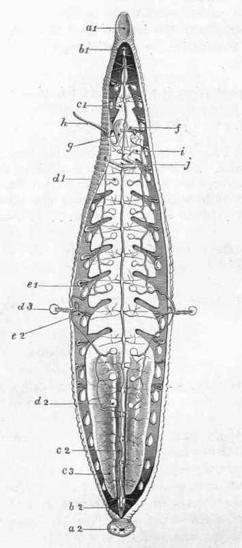
Plate XIII. Medicinal Leech. Hirudo medicinalis.
The integument is drawn as divided down the middle ventral line, from the posterior border of the buccal cavity or anterior sucker to the anterior border of the posterior sucker; two of the testes and two of the nephridia have been displaced outwards in the somite, lettered d 3, e 2; the rest of the organs have been left undisturbed in situ.
a 1. Anterior sucker.
a 2. Posterior sucker, formed by the fusion of seven distinct somites, to which as many ganglia, subsequently fused into the single posterior ganglion of the ventral chain, corresponded at one period of the animal's development. The anus is dorsal and anterior to the sucker.
b 1. Infra-oesophageal ganglion and first ganglion of ventral chain, very closely apposed to each other.
b 2. Last ganglion, the twenty-third of the ventral chain, composed of seven embryonic ganglia fused. This ganglion gives off from five to nine pairs of nerves, which are distributed to the posterior sucker. The penultimate ganglion gives off only one pair of nerves.
c 1. First lateral diverticulum of the portion of the digestive tract, which comes next after the pharynx.
c 2. 'Small intestine' of most authors, 'gastroileal' portion of digestive tube of Gratiolet, in which the haemoglobin of the blood undergoes changes. It ends posteriorly in a short ovoidal colon, which again ends in a short rectum, turned slightly upwards to the anus. The small intestine is a little dilated at its commencement in the interval between the two terminal sacculi c 3. This dilatation is much larger and more distinctly bilobed in the Horse-leech (Aulostoma Gulo).
c 3. Eleventh lateral diverticulum of right side prolonged downwards on either side of the small intestine and colon, as far as the point where the rectum begins.
d 1. The most anteriorly placed of the nine testes of either side, communicating by a short transverse duct passing outwards, with a vas deferens common to it and the eight posterior testes, and anteriorly convoluted. These convolutions are seen in this figure in the space on the right side bounded by the lines lettered i and f.
d 2. Last or ninth testis of right side.
d 3. Sixth testis, displaced outwards so as to show its connection with the vas deferens. e 1. Segmental organ or nephridium.
e 2. Segmental organ or nephridium. The testis is displaced outwards. In relation with it is a coecal process from the nephridium, the testis lobe, which is surrounded by the perinephrostomial sinus. The vesicle by which the nephridium opens externally lies just to the right of the line from the letter. See pp. 221-3, ante.
f. Ductus ejaculatorius of left side, leading from the convoluted portion of the vas deferens to the base of the prostatic body.
g. Prostatic body.
h. Muscular penis, surrounded where it passes out of the integument by a strong sphincter. This orifice lies in the second annulus of the eleventh somite (Whitman).
i. Ovarian sac of left side, carried upon one of the short oviducts. The ovarian sac of the other side is seen on the farther side of the nerve-cord, underneath which its oviduct passed.
j. Muscular vagina, in which after sexual congress the spermatophore is found. Between the vagina and the two oviducts, a common oviduct intervenes, which takes a tortuous course, and has its coils surrounded by a mass of loose tissue, composed of unicellular glands, which are probably the main agents in the secretion of the albumen which envelopes the eggs in the cocoon. The azygos character of the two generative outlets is especially noteworthy.
Continue to:
My Books

