Chapter VI. Displacements
Description
This section is from the book "Intra-Pelvic Technic OR Manipulative Surgery of the Pelvic Organs", by Percy H. Woodall, M. D., D.O.. Also available from Amazon: Intra-Pelvic Technic OR Manipulative Surgery of the Pelvic Organs.
Chapter VI. Displacements
The uterus is balanced, or in a manner floats, in its normal position in the pelvic cavity in a state of equilibrium. Under natural conditions when this state of equilibrium is disturbed it is quickly and readily regained as soon as the disturbing agency has ceased to operate. The uterus is endowed with a greater degree of mobility than any other internal organ in the body. Its position is normally altered by every respiratory act, descending with inspiration and rising with expiration. It is pushed backward as the bladder fills and forward by a full rectum and upward when both these organs are filled. Its position is changed by change of posture. It may be greatly displaced in a bimanual examination only to immediately return to its normal position which may vary considerably within certain limits. So to constitute a displacement the condition must be continuous and more or less fixed. Should the uterus become fixed or immobilized in what is called its "normal position" such a condition would be pathological. Limitation of physiological mobility is therefore one of the principal elements in a displacement.
The uterus is maintained in its normal position by a combination of agencies, no one of which is wholly sufficient, yet the failure of any one tends to upset the equilibrium and to produce a displacement. Of these agencies the chief is the pelvic floor composed of the levator ani muscles with their associated lesser muscles and fasciae. These form the pelvic diaphragm, the "pelvic sling." They are the structures closing the pelvic outlet and are the foundation support of the pelvic organs. When their function is lost displacement is almost inevitable, sooner or later.
The adjacent organs not only afford a bed upon which the uterus lightly rests when all is well, but when equilibrium is disturbed they afford some actual assistance in the maintenance of position. The bladder and anterior vaginal wall in front and below, the pressure of the intestines upon the superior surface of the fundus, the posterior vaginal wall and the rectum posteriorly are all factors in preserving position. These agencies must be in normal condition. Should they become atrophic or otherwise lose their tone they cease to provide support. Should they become congested or inflamed their additional weight would serve to disturb rather than maintain position.
The abdominal walls have an important, though an indirect, influence on uterine position. When they are in normal tone they assist in maintaining the position of the abdominal organs and prevent their descent and pressure upon the pelvic organs. That their "sustaining power" is more than this, I doubt.
The uterine ligaments possess a certain degree of elasticity, or tone, which operates in a slight degree to restore equilibrium after it is disturbed. They give but little active support to the uterus until a considerable degree of displacement has occurred.
Posture is an important factor in the maintenance of equilibrium. It operates not only through its effect on the abdominal walls, as will be mentioned later, but also by changing the plane of the pelvic brim and allowing abdominal pressure and the abdominal -organs more direct access to the pelvis. When the normal lumbar curvature is maintained the plane of the pelvic inlet approaches the perpendicular in the upright position and the uterus is snugly ensconced beneath and behind the promontory of the sacrum. Here it is fairly well protected from the influence of the intra-abdominal pressure and the weight of the abdominal organs (Fig. 4). With a slumping of the body a straightening of the lumbar curvature occurs, the plane of the pelvic inlet is made more nearly horizontal, giving an invitation, as it were, for abdominal pressure and the abdominal organs to enter the pelvis.
Abnormal size or weight of the uterus would tend to upset equilibrium, in time, even though the other elements of support were in every way normal.
Before diagnosing a displacement the condition of the bladder and rectum should be known. It is possible for a retroversion to disappear after emptying a distended bladder and for an anteposition to be removed by emptying an impacted rectum. In fact, these organs should be emptied before an examination is made.
Theoretically displacements may occur in any direction. Actually they occur most frequently in a forward and backward and a downward direction. Occasionally lateral displacements are seen. Combinations of these displacements may occur as antero-lateral, postero-lateral, or some degree of torsion with or without an accompanying displacement. Downward displacements are commonly in a backward direction also.
Of the anterior, posterior and lateral displacements two forms are described, flexions and versions. A flexion is a condition in which the uterus is bent upon itself and the angle existing between the body and the cervix is disturbed. The point of bending is usually at the junction of the body and the cervix, though it may rarely occur at some other point. Either causing or resulting from the flexion there is a diseased or weakened condition of the tissues at the point where it occurs, giving rise to a two-fold pathological condition in flexions. A version is a turning of the uterus as a whole, the angle between the body and the cervix being unchanged.
A slight degree of lateral displacement may be considered normal and is probably due to a congenital shortening of the broad ligament of one side. Some authorities claim that the uterus lies nearer the left than the right side of the pelvis. Such conditions interfere with mobility very slightly, if at all, and have no pathological significance.
An abnormal lateral displacement usually occurs as a secondary and minor accompaniment of one of the other forms of displacement. It may, however, occur alone or as the predominant disorder. In such cases the uterus may be forced toward one side of the pelvis by some form of pelvic tumor or by the swelling from some pelvic inflammation originating on the opposite side. In the latter case the uterus may be later displaced toward the side upon which the inflammation originated by the contraction of the inflammatory exudate. Adhesions resulting from such inflammatory exudate are the most common cause of lateral displacements. (Figs. 12,13,14.)

Fig. 12. Left Latero-version of the Uterus. The uterus is crowded to the left side of the pelvis, the long axis of the uterus inclines to the left. The cause of the displacement is a broad ligament cyst the right side adherent to the wall of the pelvis. (Findley, Diseases of Women.)
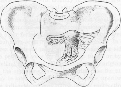
Fig:. 13. Left latero-displacement of the Uterus. The left broad ligament is thickened and contracted and has drawn the uterus to the left. (Findley. Diseases of Women.)

Fig. 14. Contraction of the left broad ligament drawing the uterus into a left lateral position. (Findley, Diseases of Women.)
Continue to:
My Books

