Tumours And Parasites Of The Liver. Continued
Description
This section is from the book "A Manual Of Pathology", by Joseph Coats, Lewis K. Sutherland. Also available from Amazon: A Manual Of Pathology.
Tumours And Parasites Of The Liver. Continued
Secondary Cancer
This is of very frequent occurrence in the liver. There may be a direct extension of a cancer from the gall-bladder or the stomach. In the latter case the organ becomes adherent and the cancer grows into the liver. There may thus be a large tumour, sometimes with a cavity communicating with an ulcerated surface in the stomach.
Metastatic cancer is more frequent. The primary tumour is usually in the stomach or intestine, but it may be in the oesophagus, uterus, mamma, or elsewhere. In most cases the cancer extends to the liver by the portal vessels (see further on as to Extension of Cancers in the Abdomen), but it may also occur by the hepatic artery in cases of cancer of the mamma and other external parts.
As the cancer is sown in every part of the liver, the consequence is the development of numerous tumours which may be.found in all stages of growth. It is usual to find a large number of isolated tumours of circular form and pale colour (see Fig. 412). They are seated in the liver tissue, but those near the surface produce rounded elevations which can often be felt through the abdominal parietes during life. Even to the naked eye the tumours show evidences of fatty degeneration in the appearance of an opaque yellow coloration. The absorption of the degenerated cells is also indicated by the partial contraction of the larger tumours in their central parts. In the superficial ones a dimpling or Umbilica-tion of the, rounded projections is commonly visible. The liver, as a whole, is sometimes enormously enlarged, weighing not infrequently as much as ten or twelve pounds.
An exceedingly rare form of secondary cancer is that represented in Fig. 413, in which there was a congeries of cysts as well as solid tumours, the walls of the cysts and the tissue of the solid tumours presenting cancerous tissue.
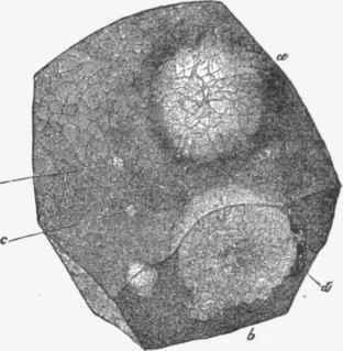
Fig. 412. - Piece of liver with secondary cancerous tumours in it. A larger one, a, is viewed from the surface. Another large one, b'in section. There are several smaller tumours, e, c; it, a vein in section. (Virchow).
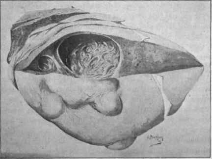
Fig. 418. - Cystic cancer of liver. (From a painting by Dr. A. Maophail).
The cysts arose by necrosis of the tissue and contained blood and debris.
In some cases the new-formation is not so much in the form of individual tumours as a general Cancerous infiltration, as if almost throughout the liver a simultaneous development had occurred, and the cancerous tissue had grown vigorously, displacing the proper hepatic tissue.
In structure the secondary tumours follow the primary ones. For the most part it is soft cancers of the stomach that form the original tumours, and we have a well-formed stroma with irregular masses of epithelial cells. But sometimes the primary tumour is an epithelioma, and in that case the tumours in the liver are usually fewer in number, firmer and more distinctly defined. When the primary tumour is in the oesophagus the structure may be that of the Plat-celled epithelioma with regular laminated capsules. When the primary tumour is in such a position that the cancer reaches the liver by the hepatic artery, the tumours are usually smaller in size and the liver not so much enlarged. This was at least very strikingly seen in a case observed by the author, where the primary tumour was in the mamma.
The cancerous material as it is brought by the blood frequently develops inside the vessels, and particularly in the portal veins and the capillaries. The growing tumours cause atrophy of the proper hepatic tissue. The hepatic cells are often to be seen at the peripheral parts of the tumours arranged in concentric layers as pressure is exercised, and in various stages of atrophy.
The numerous developing tumours press on the bile ducts and obstruct them, and this occurs all the more as the growths mostly arise by embolism of the portal vessels, and are, in the first place, related to them. As the ducts are in the immediate neighbourhood of these vessels, they are liable to be pressed on very soon. Hence General icterus is a common result, and there is also a special Pigmentation of the hepatic tissue (see ante).
The portal vessels suffer obstruction, not only by the pressure of the tumours, but as being the seat of their primary growth, and hence Ascites is a common result.
Sarcoma is of occasional occurrence in the liver in the spindle-celled and pigmented forms. Some cases of melanoid sarcoma have been described as primary tumours, but secondary tumours following melanotic sarcomas of the eye are more common. The liver may be largely occupied by these black tumours, and greatly enlarged. Here also the growth appears to occur in the blood-vessels and results in the destruction of the hepatic tissue proper.
Parasites Of The Liver
These have already been somewhat fully described. The most important constitutes the Hydatids of the liver arising from the Tsenia echinococcus. When the liver is examined after death there is found a sac or sacs of very various size, up to that of a man's head. The larger ones produce necessarily great enlargement of the liver as a whole, and atrophy of the hepatic tissue around them. They present first a connective tissue capsule, inside which the proper wall of the cyst appears. As the vesicles are sometimes much broken down, and it may even be difficult to find hooklets, it is important to remember that the proper cuticula of the cvst is lamellated. In the contents of such cysts there are frequently orange coloured masses consisting of crystals having the usual form of the haematoidin or bilirubin crystals. They are probably composed of bilirubin. The multilocular and exogenous forms of hydatids are to be remembered, the former resembling colloid , cancer in its general appearance.
It is not uncommon to meet with the remains of a dead echinococcus in the liver. It shows a capsule which contains an indefinite debris in which pieces of the chitinous membrane are visible (Fig. 414). There may be several such lesions present. In a case of this kind the author found in one of the cysts along with calcareous infiltration a production of true bone in the wall of a small collapsed cyst. In Fig. 189, p. 398, a microscopic section of a shrunken hydatid is reproduced.
The Distoma hepaticum, sinense, and lanceolatum occur in the bile ducts, the Distoma haematobium in the portal vessels. (See under Animal Parasites.) The Pentastoma denticulatum also occurs in the liver.
Literature
Nodular hyperplasia - Friedreich, Virch. Arch., xxxiii., 1865; Hoffmann, ibid., xxxix., 1867; Sabourin, Rev. de med., 1884; Simmonds, Deutsch. Arch. f. klin. Med., xxxiv., 1886; Greenish, Wien. Med. Jahresb., 1882. Prmmrg cancer - Rindfleisch, Path. Hist. (Syd. Soc), 1872-73; Greenfield, Path, trans., xxv., 1874; Mahomed, ibid., xxviii., 1877; Whipham, ibid., xxii., 1871; Paul, ibid., xxxvi., 1885; Waldeyer, Virch. Arch., lv., 1872; Perls, (Cirrhosis car-cinomatosa) ibid., lvi., 1882; Dreschfeld, ibid., and Jour, of anat. and phys., xiv., 1880; Weigert, Virch. Arch., lxvii., 1876; Hilton Fagge, Path, trans., xxviii.,1877; Pye Smith, ibid., xxxi., 1880; Hanot et Gilbert, Etudes sur les malad. du foie, 1888. Cysts - Sabourin, Arch, de physiol. norm, et path., x., 1882, and Progres med., 1884, No. 20; Becklinghausen, Virch. Arch., lxxxiv., 1881; Kennedy, Ed. Lab. Beports, iii., 177, 1891.
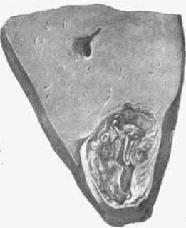
Fig. 414. - Portion of liver with collapsed hydatid cyst. Natural size.
Continue to:
My Books

