The Tunics Of The Eyeball
Description
This section is from the book "A Manual Of Physiology", by Gerald F. Yeo. Also available from Amazon: Manual Of Physiology.
The Tunics Of The Eyeball
The organ of vision of vertebrate animals is enclosed in a firm case of fibrous tissues called the sclerotic coat, which is continuous with the sheath of the optic nerve. It is seen between the eyelids under the transparent conjunctiva, and known as the white of the eye. It gives shape and protection to the eye, and though translucent, is not transparent. In front, a round, window-like portion, called the cornea, forms the anterior segment of this protecting covering of the eyeball. The cornea is distinguished from the sclerotic not only by its glass-like transparency, but also by being part of a lesser sphere than the sclerotic, and thus projecting a little more than the rest of the bulb.
Closely attached to the inside surface of the sclerotic is a soft, thin, black, vascular sheet of tissue called the choroid coat, which supplies the eyeball with blood. It is made up chiefly of blood vessels and stellate, pigmented, connective tissue cells. Its outer layer is traversed by arteries and veins of relatively large size, and its inner layer is composed of a dense network of close-meshed capillary vessels. As the cornea region is approached, the choroid is peculiarly modified and thrown into folds, called ciliary processes, forming a series of vascular folds, radiating from the margin of the cornea. At the edge of the cornea the choroid is more firmly attached to the sclerotic by a circular muscle {ciliary muscle), and also by bands of tissue from the posterior surface of the cornea, which hold it in position; the fibres of the ciliary muscle, running under the ciliary processes, radiate from the margin of the cornea toward the choroid, to which they are attached. In a modified form, known as the iris, this vascular and pigmented coat of the eye leaves the sclerotic, and hangs freely in a fluid, being recognized through the clear cornea as a colored circular curtain, attached to the inside of the periphery of the cornea, having a central aperture, which looks black, and is familiarly known as the pupil. The pupil is merely an opening in the iris, which allows the rays of light to pass into the interior of the eyeball.
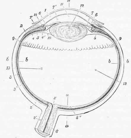
Fig. 216. Diagram of a horizontal section through the human eye.
i. Cornea; i'. Conjunctiva; 2. Sclerotic; 3. Choroid; 4. Ciliary processes; 4'. Ciliary muscle; 5. Suspensory ligament of lens; 6. So-called posterior chamber, between the iris and the lens; 7. Iris; 7'. Anterior chamber in front of the iris; 8. Optic nerve; 8'. Entrance of central artery of the retina; 8". Central depression of retina or yellow spot; 9. Anterior limit of the retina; 10. Canal of Petit in front of the hyaloid membrane; 11. Aqueous chamber; 12. Crystalline lens; 13. Vitreous humor; 14. Circular venous sinus which lies around the cornea; a - a, anterior-posterior, and b - b, transverse axis of bulb.
Besides supplying nutrition to the non-vascular central parts of the eyeball, the choroid is useful in vision by preventing the reflection of the light from the background of the eye in such a way as would cause irregularity of its distribution, and thus dazzle and interfere with the distinctness of the image. The choroid is elastic, and can move over the neighboring sclerotic; it can be drawn forward by the contraction of the radiating ciliary muscle, which acts as a tensor of the choroid membrane.

Fig.217. Showing the course of the fibres of the optic nerve, N, as they pass along the inner surface of the retina, R, to meet the ganglion cells, g, whence special communications pass outward to the layer of rods and cones in the pigment layer,p, next the choroid, c, within the sclerotic, S.
The iris has a special power of motion, by means of which the opening in it can be made smaller, so as to regulate the amount of light admitted to the eye, and cut off more or less of the rays which would pass through the margin of the dioptric media. The importance of this will be better understood further on.
Within the choroid coat, and in immediate contact with it, is the nervous coat, or retina, formed by the expansion of the optic nerve, which passes toward the sclerotic obliquely, and enters it somewhat to the nasal side of the axis of the eye. The retina lines all the back part of the eyeball, and stretching forward, becomes fused with the ciliary processes, where, however, the nervous elements of the coat are wanting. The fibrils of the optic nerve reach the inner surface of the coats of the eye, and lie in immediate relation to the transparent medium, which occupies the greater part of the bulb. The fibres then lie internally to their terminals, which turn outward and are set against the choroid coat. The ultimate nerve endings are situated in pigmented protoplasmic cells, which form the outer layer of the retina.
Continue to:
My Books

