Alimentary Canal
Description
This section is from "The American Cyclopaedia", by George Ripley And Charles A. Dana. Also available from Amazon: The New American Cyclopædia. 16 volumes complete..
Alimentary Canal
Alimentary Canal, a tubular passage, existing in man and all the higher animals, composed principally of a muscular layer and a mucous membrane, extending from the mouth to the anus, and designed for the reception, transmission, and digestion of the food or aliment. The cavity of the alimentary canal is continuous, anatomically, from its commencement to its termination, forming a hollow passage through which the food is carried in the digestive process. Its different parts are, however, partly separated from each other at various points by constrictions and muscular bands, which are alternately closed and opened, to allow of the temporary retention or onward movement of the alimentary materials. The different portions of the canal are also distinguished from each other by varieties of form and size, the development of their muscular layers, and the structure of their mucous or lining membrane. Owing to this variety of structure, and the different characters of the secretions produced, the action of the alimentary canal upon the food varies in its different parts; i and the process of digestion to which the food is subjected consists of the successive or combined operation of the whole.
The principal portions into which the canal is thus divided, in the human subject, are known as the mouth, the oesophagus, the stomach, the small intes-tine, and the large intestine. - The mouth is the cavity included between the opening of the lips in front and the fauces behind. In it are the teeth, intended for the mastication and comminution of the food; the tongue, a muscular and sensitive organ, which subserves both the sense of taste and the proper movement and ad-mixture of the food in mastication; and a lining membrane which contains mucous glandules destined to supply a viscid secretion form-ing part of the saliva. There are also the pa-rotid, submaxillary, and sublingual glands, situated in the immediate vicinity of the mouth,, which pour their secretions into its cavity, and thus complete the formation and supply of sa-liva, which is mingled with the food in mastication and reduces it to the condition of a soft, pasty mass. - Immediately behind the fauces is the pharynx, a short funnel-shaped passage leading directly to the oesophagus. The latter is a nearly straight tube of uniform size, about nine inches long and rather less than one inch in diameter.
It passes through the neck and posterior region of the chest to the upper part of the abdomen, where it terminates in the stomach. It has a double layer of transverse and longitudinal muscular fibres, by whose peristaltic or wave-like contractions the masticated food is rapidly carried from above downward. Its lining membrane is of a simple structure, and produces only a small quantity of mucus, destined by its lubricating qualities to facilitate the passage of the food. The oesophagus, in fact, is simply an organ of transmission, by which the ! food is transferred from the mouth to the stomach, where the more important digestive actions are to begin. - The stomach is a dilatation of the alimentary canal, lying transversely across the upper part of the intestine. Toward the left side it expands into a wide hemispherical sac or pouch; toward the right side it becomes narrowed to a smaller diameter, where it unites with the upper extremity of the abdomen. The orifice by which the stomach communicates with the oesophagus is called the cardia (Gr. kapδia, the heart), because it is situated near the heart; that by which it communicates with the intestine is called the pylorus (Gr. πυλωρóς, a gatekeeper). Both are provided with a special circular bundle of muscular fibres by which the food, once in the stomach, is retained there for a time, to allow of the secretion and operation of the gastric juice.
The gastric juice is secreted by the mucous membrane of the stomach, which is soft, glandular, and vascular in texture, and, when stimulated by the contact of the food, pours out the gastric juice in considerable abundance, as the perspiration is exuded by the skin. - Next the stomach follows the small intestine. This is a tubular canal about 25 feet in length and between one and two inches in diameter. It is thrown into numberless folds and convolutions, by which, notwithstanding its great length, it occupies a comparatively moderate space in the abdomen. It is attached to the abdominal portion of the spinal column by a thin, flexible membranous sheet termed the mesentery, which, while retaining it in its proper position, allows of the necessary movement of its different convolutions upon each other. Its muscular layers are well developed and active, -and by their contractions continuously urge the semi-fluid ingredients of the food through the tortuous windings of its internal cavity. Its mucous membrane is provided, first, with a great number of glandular follicles which secrete the intestinal juice, one of the active agents in digestion; and secondly, with minute filamentous vascular prominences or villi, which are so abundant and thickly set as to give its internal surface a velvety texture, and which by their absorbent action take up from the intestine the nutritious elements of the digested food.
Into the upper part of the small intestine, a few inches below the stomach, there are also discharged two accessory secretions, namely, the bile from the liver, and the pancreatic juice from the pancreas. The small intestine terminates, in the lower part of the abdomen on the right side, by a junction at right angles with the large intestine. - The large intestine, so called from its greater capacity as indicated by a transverse measurement, is about five feet long, and from 1 1/2- to 2 1/2 inches in diameter. It extends from its commencement in the right iliac region (see Abdomen) upward on the right side of the abdomen, then transversely across to the left side, then downward upon the left side, then through an S-like convolution to the top of the pelvis, and finally through the cavity of the pelvis to the anus. At the point of junction of the small with the large intestine there are two parallel folds of mucous membrane, with their edges turned toward the cavity of the large intestine, which act as a double valve (called the ileo-caecal valve), allowing the passage of materials in this direction, but preventing their regurgitation from the large into the small intestine.
The mucous membrane of the large intestine has no villi, but is provided with simple glandular follicles, which secrete various exertmen-titious materials. This portion of the alimentary canal contains also the refuse portions of the food, which, together with the excremen-titious matters supplied by its lining membrane, assume a faecal consistency and appearance from the situation of the ileo-caecal valve downward, and are finally discharged from the lower extremity of the large intestine.
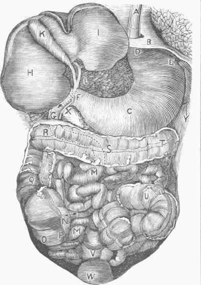
Abdominal Portion of the Alimentary Canal.
A, oesophagus; B. diaphragm; C, stomach; D, cardiac extremity of the stomach; E, great pouch; F, pylorus; G, duodenum, H, right lobe of liver; I. left lobe of liver; K, gall bladder; L, bile duct; M. small intestine; N, entrance of small intestine into the large intestine; O, caecum; P, appendix vermiformis; Q. ascending colon; R S T, transverse colon; U, sigmoid flexure; V, rectum; W, urinary bladder; X, pancreas; Y, spleen.
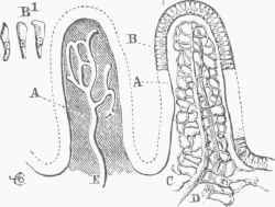
Two Villi of the Small Intestines.
A, substance of the villus; B, its epithelium, of which some cells are seen detached at B1; (J I), the artery and vein, with their connecting capillary network, which envelopes and hides the lacteal radicle. E, which occupies the centre of the villus and opens into a network of lacteal vessels at its base.
Continue to:
My Books

