The Mamma Or Breast
Description
This section is from the book "Applied Anatomy: The Construction Of The Human Body", by Gwilym G. Davis. Also available from Amazon: Applied anatomy: The construction of the human body.
The Mamma Or Breast
The name mammary gland is often given to the breast, yet the latter is composed not only of glandular tissue but also of fibrous and fatty tissue, with the usual vessels, nerves, and lymphatics. In the male the glandular portion is undeveloped, the fat is relatively scanty, and the breast as a whole is insignificant and flat. In the virgin female adult it is more spheroidal. Above the nipple it is flattened and below it is rounded. Its general shape is circular and it covers the chest-wall from the upper border of the third rib to the sixth interspace. Laterally it reaches internally almost to the sternum and externally it overlaps the edge of the pectoralis major. It lies imbedded in the superficial fascia. In its development it is simply a modified sebaceous gland. Beginning by a finger-like growth from the skin it burrows its way into the superficial fascia. It becomes compound and sends its branches in various directions, especially does it extend deeper until finally it pushes away most of the fat and rests on the fascia covering the pectoralis major muscle. This is why we find almost no adipose tissue beneath the gland but mostly between the glandular structure and the skin and around its edges. The shape of the virgin breast is due mainly to its adipose tissue and not to its glandular structure. In those who have borne children the breasts become enlarged, lax, and pendulous. After lactation is completed they again retract but rarely regain their former shape. During lactation the fatty portion of the breast may disappear and leave it apparently in a shriveled condition, yet such a breast may be functionally quite active. Therefore the size of the breast is no criterion of its milk-producing powers.
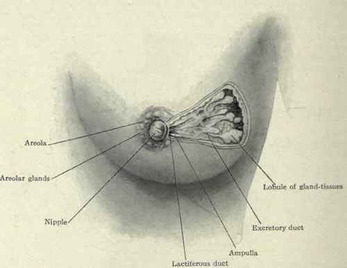
Fig. 205. - The secreting structure of the breast. (Piersol).
The secreting structure, racemose in character, is divided into ten to sixteen lobules, each of which has its duct. These lactiferous ducts begin in the acini and end in the nipple. Beneath the nipple they are dilated, each forming a sinus or ampulla. While the shape of the breast is regular in its outline the glandular tissue is not so. It possesses three projections or cusps. One of these projects inward nearly or quite to the sternum, while the other two project toward the axilla and side, one being lower than the other. These are the most common directions in which the glandular tissue is prolonged, but it may extend farther than usual in any direction; hence the wide incisions made in cases of carcinoma.
According to H. J. Stiles (Ed. Med. Jonrn., 1892, p. 1099), the secreting structure may extend posteriorly into the retromammary tissue between the layers of the pectoral fascia. Anteriorly it is prolonged with the fibrous tissue (ligaments of Cooper) almost to the skin.
The nipple, located below and to the inner side of the centre of the gland, has connected with it some circular and longitudinal unstriped muscular fibres. The longitudinal ones are attached to the lactiferous ducts and serve to retract the nipple, the circular ones to erect it. Surrounding the nipple is the areola. It is pink in the virgin and about 2.5 cm. in diameter. After pregnancy its hue becomes brownish. The tubercles of Montgomery are the numerous elevations found on the areola. They are more or less modified sebaceous glands and enlarge during pregnancy. As they secrete a milky fluid, they are often regarded as accessory milk duets. There is no fat in the nipple or areola.
The fibrous structure of the gland envelops the adipose and glandular tissue. It is simply a continuation of the fibrous septa of the superficial fascia. These septa are attached to the skin above, envelop and pass between the fatty and glandular lobules, and form a thin covering for the under surface of the gland. The breast is sometimes spoken of as having a capsule, but that simply refers to the fibrous tissue just described. This fibrous tissue follows largely the ducts, hence when affected with carcinoma it contracts and draws the nipple in. This forms the retracted nipple of that disease. The fibres that go to the skin have been named the ligaments of Cooper. The fibrous tissue forms a net-work in the meshes of which are packed the glandular structure and fat-lobules. It is this which gives the firmness and shape to the virgin breast. In lactation, the fibrous tissue softens and stretches to accommodate the increase in the glandular structure and this, with the loss of fat, causes the breast to become lax and pendulous. In palpating a normal breast between the fingers and the thumb, this firmness may feel like a foreign growth; hence this method of examination is not to be relied on. A better way is to have the patient recline, and lay the fingers flat on the breast, compressing it on the chest-wall beneath. This flattens the glandular structure and any mass can be more surely detected.
The fibrous tissue between the glandular structure and the pectoralis beneath is quite thin and loose, with large spaces in it which have been called the sub-mammary bursa. Pus readily spreads in this loose submammary tissue, but in the gland itself only with difficulty.
Continue to:
My Books

