Pathology Of The Blood. Anemia
Description
This section is from the book "A Manual Of Pathology", by Guthrie McConnell. Also available from Amazon: A Manual Of Pathology.
Pathology Of The Blood. Anemia
Primary Anemia
Primary anemias are those conditions of decreased hemoglobin and red cells in which no demonstrable cause can be found for the blood change.
Chlorosis is a form of anemia occurring mainly in girls at the age of adolescence. It is characterized by a great reduction in the amount of hemoglobin without a corresponding reduction in the number of erythrocytes. The color-index is low, frequently falls to 0.5.
The cause is unknown. Various conditions such as hypoplasia of the arterial system and of the genitalia, intestinal autointoxication, vasomotor neuroses, heredity, poor hygiene, etc., have been advanced as explanations of the condition. Unsanitary conditions about the age of puberty undoubtedly play an important part.
The blood shows certain characteristic conditions, it is very pale on account of the reduction of the hemoglobin, 40 to 30 per cent, of the normal not being unusual, sometimes it may drop as low as 20 per cent, with a color index of 0.5. The number of red cells is reduced, as a rule not below 4,000,000 but sometimes getting down to 2,000,000. Such a low count generally indicates some complication. The red cells are usually smaller than normal, very pale in the center and frequently show poikilocytosis. Nucleated red cells rarely appear, when they do the normoblasts is the common type, although megaloblasts have been found. The specific gravity is reduced in proportion to the decrease of hemoglobin, it may reach 1028.
Changes in the leukocytes in size and number are unusual.
Pernicious Anemia
Pernicious Anemia is a disease of the blood and blood-forming organs, characterized by excessive destruction associated with defective production of red cells. The amount of hemoglobin is diminished greatly at the same time but the amount in each red cell is usually greater than normal, thus giving a high color index, more frequently above one than below.
The cause of this condition is unknown although in some instances there has been an infection by intestinal parasites, particularly the Dibothriocephalus latus and the Uncinaria duodenalis. A similar blood picture has also been found as a result of numerous hemorrhages, of gastric carcinoma, and of atrophy of the gastric mucosa. The destruction of the blood may take place chiefly in the portal circulation and especially in the spleen as the result of toxic substances absorbed from the intestine. It has also been noted that certain anerobic bacteria found in the large intestine produce a substance that has marked hemolytic properties. This penetrates the wall of the intestine and enters the portal circulation.
The blood shows a marked diminution of erythrocytes often as low as 1,000,000 or even lower, 138,000. The hemoglobin is considerably reduced, 25 to 35 per cent, being common, but the relative amount in each cell is high.
There are also decided variations in the size of the red cells, megalocytes being very numerous; microcytes are also common. Nucleated red cells also occur in large numbers, the megalo-blasts usually outnumbering the normoblasts. This preponderance of the megaloblasts is practically pathognomonic of this type of anemia. The nuclei are frequently degenerated and generally show polychromatophilic degeneration. Poikilocytes are very common, numerous, and show a great variety of shapes.
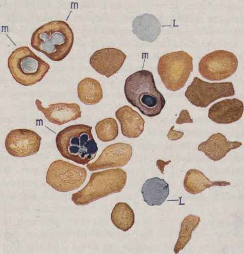
Fig. 127. - Pernicious Anemia (Cabot).
L, L, Lymphocytes; m, m, m, m, megaloblasts; cover-slips stained with Ehrlich's triacid, and drawn with camera lucida.
Coagulation is slow, specific gravity is low, 1.028, and there is little tendency to form rouleaux.
The leukocytes are generally somewhat decreased in number, averaging about 4000. This condition is practically never present in the secondary anemias. An increase in number indicates, as a rule, some complication. The decrease is mainly in the polymorphonuclear type and consequently there is a relative increase in the number of lymphocytes. A few myelocytes may be found.
On account of the extensive destruction of the red cells with the consequent liberation and breaking down of the hemoglobin there is an extensive deposit of iron in the liver, spleen, marrow and other organs. On account of the lessened oxygen the chief lesion of the tissues is an extreme fatty degeneration of the viscera, particularly the liver, kidneys and heart muscle. The marrow of the long bones is no longer yellow, but red, and soft, and frequently shows areas of hemorrhage. Under the microscope it will be noted that the normal nucleated red cells are in part replaced by an excessive number of megaloblasts, large nucleated red cells. There is also a greatly decreased amount of fat in the marrow. In the spinal cord degeneration of the posterior columns has been described and is thought to be due to minute hemorrhages.
Splenic Anemia
Splenic Anemia is a form of chronic anemia characterized by an idiopathic enlargement of the spleen without any involvement of the lymph nodes. In Banti's disease there is the primary increase in size of the spleen with a secondary interlobular (portal) cirrhosis of the liver.
The blood shows a relatively high red cell count, a marked reduction in the percentage of hemoglobin, and a low color index. The white cells are usually decreased, but there is a relative lymphocytosis.
In the Gaucher type of splenomegaly there is a wide-spread proliferation of the endothelial lining of the splenic sinuses. The blood may contain large numbers of nucleated reds, with a high leukocytosis, and a small percentage of myelocytes. This disease usually occurs in children.
Pseudoleukemia Infantum
Pseudoleukemia Infantum is a rare form of anemia seen in children and is characterized by an enlargement of the spleen, liver, and lymph nodes. The red cells are greatly reduced, to about 1,000,000, nucleated erythrocytes are common, hemoglobin is diminished, and the leukocytes increased to 20,000 to 50,000. This latter vary greatly in shape and frequently are much larger than normal.
Secondary Anemia
Secondary Anemia is one in which there can be found an underlying cause to explain the changes present in the blood. The hemoglobin is regularly diminished to a greater extent than the red cells; it may fall as low as 15 to 20 per cent. The red cells seldom fall below one million. In severe cases normoblasts may be numerous. The size of the red cells is slightly below the normal, and there is usually some poikilocytosis. As a rule there is not much change in the leukocytes unless suppurative processes are present. The specific gravity is reduced in direct proportion to the loss of hemoglobin. The rapidity of coagulation is commonly increased.
The more common causes are acute and chronic hemorrhage, inanition, intestinal parasites, fever and certain poisons such as alcohol, lead, acetanilid and others that cause hemolysis with hemoglobinemia.
Continue to:
My Books

