Rotifera (Ehrenberg). Part 6
Description
This section is from the book "General Outline Of The Organization Of The Animal Kingdom, And Manual Of Comparative Anatomy", by Thomas Rymer Jones. Also available from Amazon: A General Outline of the Animal Kingdom and Manual of Comparative Anatomy.
Rotifera (Ehrenberg). Part 6
* Quarterly Journal of Microscopical Science, no. 1. p. 9. 1 Williamson, loc. cit.
(1147). In addition to the elaborate organization described above, the Prussian naturalist conceived that he had discovered a vascular apparatus, consisting of transverse vessels (fig. 230, n n), in which he supposed a circulation of the nutritive fluids occurred. But the vascular character of the transverse striae visible in this position is more than doubtful, as there seems every reason to suppose that the appearance depicted in the figure is due to the existence of the transverse muscular bands whereby the extrusion of the rotatory apparatus is effected, analogous to those occupying a similar situation in the Bryozoa.
1148.) The mode in which respiration is effected, in the class of animals under consideration, has been a subject of much dispute. Some have supposed the contact of water, applied to the general surface of the body, sufficient for the aeration of the nutritious juices, especially as its constant renewal would be ensured by the ciliary movements. Bory St. Vincent*, on the contrary, regarded the rotatory cilia as real gills, resembling those of fishes; and, mistaking the movements of the gizzard for the contractions of a heart, conceived these animalcules to be even superior to insects in the organization of their vascular system. Ehren-berg, however, thinks that he has discovered an internal respiratory apparatus of a most extraordinary description. In Notommata centrura he remarked seven vibrating points on one side, and six on the other, attached to two long and undulating viscera, which he elsewhere describes as being the testes of the animal (fig. 225, I); the above-mentioned points were never at rest, and appeared to be placed in determinate positions opposite to each other. Accurate observations, he says, have shown each to be a peculiar little organ, provided with a tail resembling that of a note in music, and to be thrown into vibration by three little vesicles, or folds of their inflated extremity; these organs floated freely in the abdominal cavity by their enlarged portion, while by their tail they were attached to the long tubular organ above referred to.
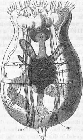
Fig. 230. Notommata clavulata (after Ehrenberg): a, rotatory organs; b, gizzard; c, stomach; d d, intestine; e, eaecal appendages to stomach; f, ovary; g, contractile vesicle; h h, lateral tubes, to which are appended the vibratory organs; n n, transverse bands, supposed by Ehrenberg to belong to a vascular system.
* Diet, des Sci. Nat., art. "Rotifera".
(1149). Ehrenberg's first idea on seeing these organs was that they formed a vascular system, executing movements of pulsation; but he now considers them as internal branchiae, or organs of respiration, to which the external water is freely admitted in the following manner: -
(1150). In many species of the Rotifera, we find, projecting from the neck of the animal, a horny tubular organ, called by Ehrenberg the calcar or spur (fig. 225, d): this he at first considered to be the male organ of sexual excitement; but he now regards it as a siphon, or a tube of respiration, through which the circumambient water passes freely into the cavity of the body. He thinks, moreover, that the periodical transparency, and the alternate distention and collapse of the animal, seen to occur regularly in almost all the Rotifera, are produced by the introduction of water into the visceral cavity and its subsequent expulsion therefrom, upon which action the fluctuations observed in the interior of the body would therefore depend. The supposition that water is injected in this manner into the body seems to be favoured by other appearances: for when the internal cavity is thus filled, all the viscera appear isolated, so that the boundaries of each can be distinctly seen; but when the water is discharged, they approximate each other, their limits become confounded, and the external membrane of the body assumes a crumpled appearance.
(1151). Upon reviewing the above account of the mode of respiration in the Rotifera, we must say that we consider that the office assigned to the little organs called branchiae is extremely problematical, especially as we have but the most vague intimations concerning the existence of a circulating system at all, much less of such a double circulation carried on in arteries and veins as the presence of such organs would infer. "I presume," says Ehrenberg, "that the branchiae possess a vascular system; for when the local contractions occur in the body of the animal, we see distinctly a certain number of filaments (vessels?) loose and delicate." The opinions of the Professor himself concerning the nature of the organs which he describes being so indefinite, we must pause before adopting the physiological views to which their admission would lead - more especially as, from the very fact of the whole visceral cavity being perpetually filled with aerated fluid, the existence of any localized organs of respiration could hardly be esteemed necessary.
(1152). The two lateral bands above mentioned (fig. 225, l), with which are connected the "trembling gill-like organs" of Ehrenberg, are now considered as constituting a peculiar apparatus, distinguished as the "water-vascular system," of which, as they exist in Lacinularia socialis, the following description is given by Professor Huxley*. In this species there is no contractile sac as in other genera; but two very delicate vessels, about 1/4000th of an inch in diameter, clear and colourless, arise by a common origin upon the dorsal side of the intestine. The vessels separate, and one runs up on each side of the body in the direction of the mouth. Arrived at the level of the pharyngeal bulb, each vessel divides into three branches: one passes over the pharynx and in front of the pharyngeal bulb, and unites with its fellow of the opposite side; while the other two pass, one inwards and the other outwards, in the space between the two layers of the trochal disk, and there terminate as caeca. Besides these, there seemed sometimes to be another branch just below the pancreatic sacs.
Continue to:
My Books

