Tibia Or Second Thigh (Fig. 311)
Description
This section is from the book "The Horse - Its Treatment In Health And Disease", by J. Wortley Axe. Also available from Amazon: The Horse. Its Treatment In Health And Disease.
Tibia Or Second Thigh (Fig. 311)
A long bone extending from the femur to the hock joint. It is broad above and narrow below. The superior extremity articulates with the condyles of the femur, and is divided into two lateral articular portions by a conical projection (tibial spine). In front, and extending for some distance down the bone, is a projecting ridge, inclining somewhat outward; this is known as the "tibial crest". On the outer side of the head of this bone above, a small smooth space is noticed for articulation with the fibula.
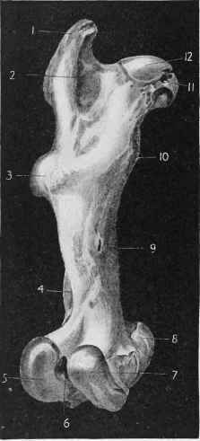
Fig. 309. - Os Femoris (Posterior Aspect).
1 Great Trochanter. 2 Trochanteric Fossa.
3 Trochanter Minor Externus. 4 Supra-condyloid Fossa. 3 External Condyle. 6 Inter-condyloid Fossa. 7 Internal Condyle. 8 Internal Trochlea. 9 Nutritive Foramen. 10 Internal Trochanter.
11 Fossa for attachment of Ligamentum Teres.
12 Head of Femur.
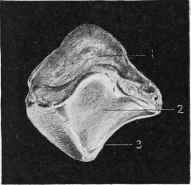
Fig. 310. - Patella (Superior and Posterior Face).
1 Superior Face. 2 Articular Face. 3 External Border.
The lower extremity of the bone, smaller than the upper, presents two deep grooves and three prominent ridges which are covered with cartilage and articulate with the astragalus to form the "true hock joint".
The inner and outer ridge each bears a projection distinguished as the internal and external malleolus of the tibia. The former is very prominent, so much so, sometimes, as to give the inner and upper part of the hock an abnormal appearance. These projections afford attachment for strong-connecting ligaments uniting the bones of the hock joint.
The articular grooves, which they assist in forming, take an oblique direction from behind outward and forward.
Continue to:
My Books

