The Thigh, Anatomy, Mass, Below The Knee, Below The Knee, The Knee
Description
This section is from the book "Constructive Anatomy", by George B. Bridgman. Also available from Amazon: Constructive Anatomy.
The Thigh, Anatomy, Mass, Below The Knee, Below The Knee, The Knee
The Thigh
Anatomy
From the head of the femur (trochanter) to the outside of the knee runs a band of tendon called the ilio-tibial band. It makes a straight line from the head of the thigh bone to the outside of the knee.
The rectus femoris muscle makes a slightly bulging straight line from just below the iliac crest to the knee cap.
On either side of the latter is a twin mass of muscles. That of the outside (vastus externus) makes one mass with it, and slightly overhangs the ilio-tibial band outside. That of the inside (vastus interims) bulges only in the lower third of the thigh, and overhangs the knee on the inside.
Behind and inside of this is the groove of the thigh occupied by the sartorius muscle, passing from the ilium above to the back of the knee below.
Behind the groove is the heavy mass of the adductors, reaching two-thirds of the way down the thigh.
Behind groove and adductors, around the back of the thigh and to the ilio-tibial band outside, is the mass of the ham-string muscles, whose tendons are found on either side of the knee at the back. It is a dual mass of muscle, dividing above the diamond-shaped popliteal space at the back of the knee, whose lower corner is formed by the gastrocnemius muscle, similarly divided.
Mass
The mass of the thigh is inclined inward from hip to knee, and is slightly beveled toward the knee from front, back and outside.

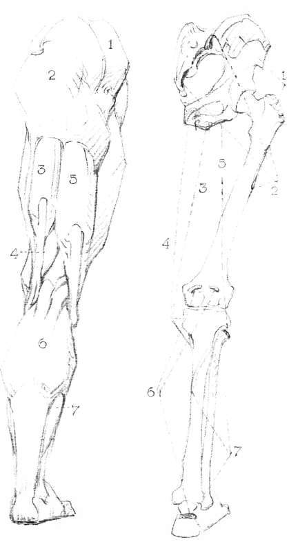
Lower Limbs. Bones of the Lower Limb.
Hip - Pelvis.
Thigh - Femur.
Leg - Tibia and Fibula (outside).
Muscles of the Lower Limb, front view:
1 Tensor of the fascia lata.
2 Sartorius.
3 Rectus femoris.
4 Vastus externus.
5 Vastus internus.
6 Tibialis anticus. 7 Peroneus longus.
8 Extensor longus digitorum.
Tensor Vaginae Femoris (Tensor Fasciae Femoris)
From crest of ilium, front end, to fascia lata, or ilio-tibial band.
Action
Tenses fascia and rotates inward thigh.
Sartorius
From spine to ilium in front to tibia inside.
Action
Flexes, abducts and rotates inward thigh.
Rectus Femoris
From anterior inferior spine of ilium to common tendon of patella.
Action
Extends leg.
Vastus Externus
From outer side of femur to common tendon of patella.
Action
Extends and rotates outward leg.
Vastus Internus
From inner side of femur to common tendon of patella.
Action
Extends and rotates inward leg.
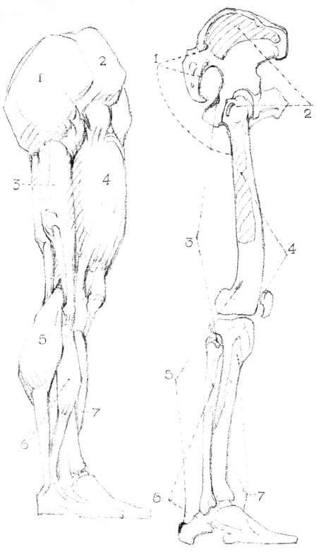
Lower Limbs. Muscles of the Lower Limb, back view: 1 Gluteus medius.
2 Gluteus maximus.
3 Semi-tendinosus.
4 Semi-membranosus.
5 Biceps femoris.
6 Gastrocnemius.
7 Soleus.
Gluteus Medius Gluteus Maximus
Page 176
Semi-Tendinosus
From ischial tuberosity to tibia.
Action
Flexes knee and rotates inward leg.
Semi-Membranosus
From ischial tuberositytotibia.
Action
Flexes knee and rotates leg inward.
Biceps Femoris
Long head from ischial tuberosity; short head from femur, to head of fibula.
Action
Flexes knee and rotates thigh outward.
Gastrocnemius, page 188. Soleus, page 192.

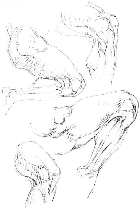
Lower Limbs. Knee Joint, back view.
Ham-Strings, Gastrocnemius and Popliteal Space.
Lower Limbs. Muscles of the Lower Limb, outer view: 1 Gluteus maximus.
2 Gluteus medius.
3 Biceps femoris.
4 Vastus externus.
5 Gastrocnemius.
6 Peroneus longus.
7 Tibialis anticus.
Below The Knee
Gastrocnemius
From tuberosities of femur to tendon of Achilles.
Action
Extends foot, raises body in walking".
Peroneus Longus
From head and upper part of fibula passes beneath foot from outside, to base of big toe.
Action
Extends ankle and raises outer side of foot.
Tibialis Anticus
From upper and outer two-thirds of tibia to inner side of foot.
Action
Flexes ankle and raises inner side of foot.

Lower Limbs. Muscles of the Lower Limb, inner view.
1 Rectus femoris.
2 Vastus interims.
3 Sartorius.
4 Gracilis.
5 Semi-tendinosus.
6 Semi-membranosus.
7 Gastrocnemius.
8 Soleus.
Below The Knee
Soleus
From upper part of fibula and back of tibia to tendon of Achilles.
Action
Extends foot and lifts body in walking'.
Extensor Communis Digitorum (Extensor Longus Digitorum Pedis)
From tibia and front of fibula to second and third phalanges of toes.
Action
Extends toes. Diagram, page 183.
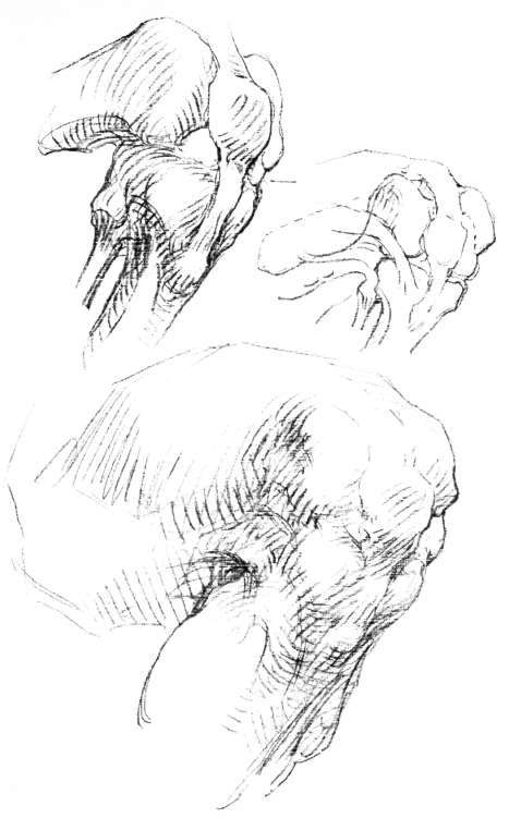
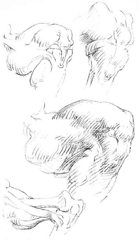
The Knee
The knee must be thought of as a square with sides beveled forward, slightly hollowed at the back, and carrying in front the knee cap; like the stopper of an ink well.
A powerful ligament connects the cap with the high ridge of the shin bone below, the two sliding together on the end of the thigh bone, which is therefore exposed in flexion. The cap is always at the apex of the angle made by thigh and leg.
From the knee cap at the top rise the three muscles already described, the rectus by a tendon narrowing upward; the vastus externus by a tendon angling slightly out: the vastus interinus bulging" prominently from the corner of the cap.
When the knee is straight, its bursa, or water mattress, forms a bulge on either side in the corner between the cap and its tendon, exactly opposite the joint itself; the knee cap being always above the level of the joint.
The back, when bent, is hollowed out by the hamstring" tendons on either side; when straight, the bone becomes prominent between them, making, with these tendons, three knobs.
The inside of the knee is larger; the knee as a whole is bent convex toward its fellow. The hip socket, the knee and the ankle are all in line: but the shaft of the thigh bone is carried some distance out by a long neck, so that the thigh is set at an angle with the leg.
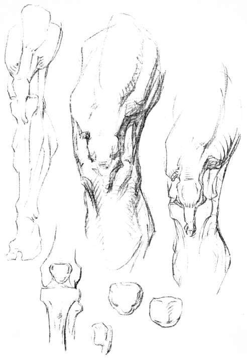
The Knee. Knee, outer view.

The Knee. Knee.
1 Pad or sack.
2 Common tendon.
3 Patella or knee-pan.
4 Ligament of the patella.
The Knee. Knee, inner view.
Continue to:
My Books

