The Thorax
Description
This section is from the book "Applied Anatomy: The Construction Of The Human Body", by Gwilym G. Davis. Also available from Amazon: Applied anatomy: The construction of the human body.
The Thorax
The thorax or chest is that portion of the trunk which lies between the neck and the abdomen. It is composed of a bony framework reinforced by soft parts, and contains the main organs of circulation and respiration. The oesophagus, an organ of the digestive tract, simply passes through it to the regions below. The chest-walls as well as the parts contained within them are affected by wounds and disease, especially the heart and its associated great vessels, and the lungs and pleurae. These organs are essential to life, like the brain and spinal cord, and like them, are encased in a bony framework. It is an example of bones performing a protecting function in addition to a supporting one.
The functions of the heart and lungs are influenced by constitutional diseases in addition to their own local affections, hence they serve as guides to the general bodily condition, and the condition of the respiration and circulation is continually being examined for the purposes of diagnosis, prognosis, and treatment, even when the heart and lungs themselves are not involved. To make these examinations intelligently, necessitates a knowledge of the organs themselves and their relation to one another and the surrounding parts. This is essential for the physician even more than the surgeon.
The chest-walls are composed of a bony framework joined and bound together and covered by soft parts.
The bones of the chest consist of the sternum, rids, and thoracic vertebra. The clavicle and scapula compose the shoulder-girdle and belong to the upper extremity. The human skeleton is divided into an axial portion and an appendicular portion. The axial portion embraces the skull, the vertebral column, including the sacrum and coccyx, the hyoid bone, the sternum, and the ribs. The appendicular portion consists of the shoulder-girdles and upper extremities and the pelvic girdles and lower extremities.
The bony chest is subject to disease and injury as well as to defects in development, and to deformities due to these causes.
Shape Of The Chest
The chest is conical in shape, being small above and large below. In transverse section it is kidney-shaped, the hilus of the kidney being represented by the vertebrae. In the foetus the anteroposterior diameter is greater than the transverse, thus resembling the thorax in the lower animals. After birth and in infancy the two diameters are nearly equal, hence we have the rounded chest of the child. As growth and development progress the transverse diameter increases more than the anteroposterior, so that at about the second year the chest has become oval and in adults the transverse diameter is one-fourth greater than is the anteroposterior.
Variations in the shape of the chest are mainly the result of disease. In childhood, rachitic disease (rickets) produces a lateral flattening and a projection of the sternum If the sternum projects markedly it constitutes what is known as pigeon breast, the chest in such a condition being longer from before backward than from side to side. In this disease also there may be a depression on each side of the sternum, the back is rounded owing to the bending of the vertebral column, and the points of junction of the ribs and cartilages are enlarged, this latter constituting what is known as beading of the ribs. These beads are felt as rounded enlargements at the sternal extremities of the ribs and form a line parallel to the sternum above and sloping outward below. This line of beads has been called the "rachitic rosary." From the level of the ensiform cartilage a groove passes out toward the sides; this has been called "Harrison':s groove" (see Fig. 193). Sometimes the lower end of the sternum is pressed inward, forming a deep funnel-shaped depression constituting the deformity known as "funnel chest" or the "Trichterbrust" of the Germans. This condition of the chest, with the exception of the beading, is also produced in children by obstruction to the breathing from enlargement of the tonsils, from the presence of adenoid growths in the pharynx, and from hypertrophy of the turbinate bones, all of which interfere particularly with nasal respiration.
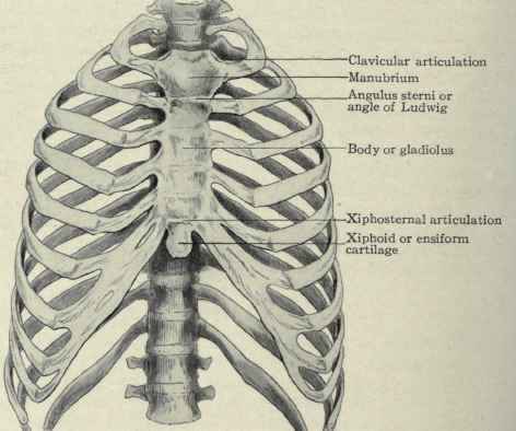
Fig. 192. - The bony thorax.
Diseases of the lungs and pleurae alter the shape of the chest. In emphysema and when distended by plural effusions, the thorax becomes more rounded in shape, forming what is called the "barrel-shaped chest." In phthisis the wasting of the tissues contraction of the lungs causes the chest to collapse. The ribs slope more sharply downward and the chest becomes longer and flatter, the anteroposterior diameter being diminished. The angle made by the lower ribs as they ascend to the sternum is called the costal angle; this becomes decreased in phthisis. This form of chest is known as the ' "phthisical chest".

Fig. 193. - Child showing Harrison's groove opposite the ensiform cartilage.

Fig. 194. - Barrel chest of emphysema.

Fig. 195. - Flat chest of phthisis.
When the flatness is marked it is sometimes called the "flat chest." When the scapulae project like wings it is called "alar" or "pterygoid chest".
In Pott's disease, or caries of the spine, as the kyphosis develops the chest falls forward and its anteroposterior diameter is increased. The abdominal contents are crowded up into the chest and push the sternum and lower ribs forward. Associated with this deformity is oftentimes a lateral deviation of the parts above the site of the disease.

Fig. 196. - Kyphosis from Pott's disease. or caries of the lower thoracic vertebrae. The curvature is an angular anteroposterior one.
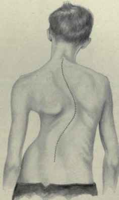
Fig. 197. - Scoliosis, or lateral curvature of the spine.
In scoliosis, or lateral curvature of the spine, the distortion is uneven, being a compression of the thorax from above downward and a twisting around a vertical axis. The deformity is frequently so severe as to cause the lower ribs to rest on the iliac crests. It is in order to detect these diseases in their early stages that a knowledge of the shape of the normal chest is so essential.
Continue to:
My Books

