Section VIII The Respiratory Organs
Description
This section is from the book "Handbook Of Anatomy For Students Of Massage", by Margaret E. Bjorkegren. Also available from Amazon: Handbook Of Anatomy For Students Of Massage.
Section VIII The Respiratory Organs
The Organs of Respiration are the lungs and trachea, the latter being the passage by means of which air is carried from the pharynx to the lungs.
Breathing consists of the acts of inspiration and expiration; in the former, air is drawn into the pharynx through either the nose or mouth and conveyed by means of the trachea to the lungs; the air is expelled in the same way.
The upper part of the air passage, the larynx, is specially modified by cartilages and muscles to produce sounds- i.e., the voice- during expiration.
Pharynx
The Pharynx is a large space behind the nose and mouth in the lower part of which are the openings of the larynx and oesophagus; the pharynx is compressed laterally, and its anterior wall is practically non-existent, the lateral walls being attached to the sides of the nasal, buccal and laryngeal orifices. The posterior wall is attached by areolar tissue to the muscles in front of the first six cervical vertebrae, and above, it is attached to the basilar process of the occipital bone and to the temporal bones.
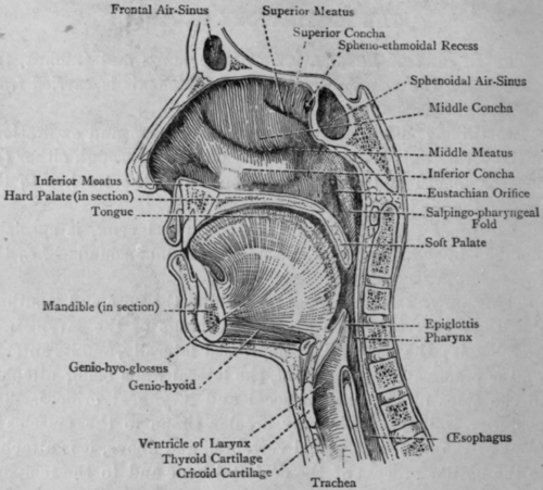
Fig. 51. - The Pharynx.
The upper part of the pharynx is almost separated off from the lower part by the soft palate, which projects backwards from the palatal processes of the maxillary bones, and in this upper part is found the orifices of the Eustachian tube and the pharyngeal tonsil. Below the soft palate is the tonsil on each side. Below this the pharynx rapidly narrows as it passss the opening of the larynx and becomes the oesophagus.
Larynx
The Larynx is the upper part of the air passage, and is placed in front of the fourth, fifth, and sixth cervical vertebrae. It consists of several cartilages held together by muscles. The largest-the thyroid cartilage-cousists of two large plates of cartilage joined at an angle in the middle line; in the male this angle is about 90 degrees, and projects forward, forming what is called the "Adam's apple." Below the thyroid cartilage is the cricoid cartilage, in shape like a signet ring with the narrow part in front. The interval between the two can easily be felt in the living subject. The thyroid cartilage is joined by a strong membrane to the hyoid bone, and from its inner side the epiglottis, a cartilaginous process, projects upwards to the back of the tongue.
Trachea
The Trachea is the continuation of the air passage; it begins just below the cricoid cartilage at the level of the sixth cervical vertebra and ends at the fourth thoracic vertebra by dividing into two bronchi. The trachea is a muscular tube kept permanently patent by rings of cartilage, which, however, are not complete posteriorly, so the organ is not quite cylindrical. These cartilaginous rings are continued in the bronchi.
The trachea follows the curve of the vertebral column, so passes obliquely backwards as it descends. It is in the middle line until the bifurcation is reached, where it lies slightly to the right.
Bronchi
The Bronchi pass obliquely downwards aud outwards from the bifurcation of the trachea to the roots of the lungs. The right bronchus is shorter and wider than the left, and is not so obliquely placed. The bronchi have several branches passing to the different lobes of the lungs.
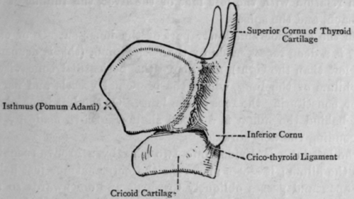
Fig. 52. - The Laryngeal Cartilages.
Continue to:
My Books

