5. Healing Of Phthisis
Description
This section is from the book "A Manual Of Pathology", by Joseph Coats, Lewis K. Sutherland. Also available from Amazon: A Manual Of Pathology.
5. Healing Of Phthisis
It is to be remembered that tuberculosis is due to an infective material, which usually goes on reproducing itself. In the healing of tubercular lesions generally there are two methods which may be followed, and in phthisis pulmonalis we have examples of each of these. On the one hand, the infective matter may be cleared out and the parts around become the seat of simple inflammatory processes, or, on the other hand, the caseous matter may have its infective character overcome and be left as a piece of innocuous dead matter in the tissues. In either case, so far as the lung is concerned, there is implied an increase in the vigour of the parts, so that the infective character of the matter may be annulled.
When cavities have formed by the softening of the caseous matter, the disease may pause. In that case the wall of the cavity comes to be composed of healthy granulation tissue, which develops into connective tissue as in the ordinary cicatrix. If the cavity is so situated that contraction can occur, then there may be a shrinking till it is completely obliterated, and a Cicatrix takes its place. On the other hand, circumstances may allow of only partial contraction, and the cavity remains, but its wall comes to be formed of connective tissue without any trace of recent tuberculosis (see Fig. 374).
In other cases the caseous matter fails to soften, the disease is checked before cavities are formed, and in that case we have necrosed structures lying in the lung. In this case granulation tissue is formed around the dead matter, and may partly eat into it. By development into connective tissue a capsule is formed around the dead matter, and the latter by and by becomes impregnated with lime salts, so that ultimately particles of lime or considerable pieces of cretaceous matter are present in the midst of a cicatrix.
It is worthy of note that the connective tissue formed in both these forms of healing is deeply pigmented, so that in this respect the processes are comparable to those concerned in fibroid phthisis.
Healing by one or other of these processes is of frequent occurrence. In the course of post-mortem examinations one very frequently meets with pigmented cicatrices, often with chalky particles in their midst, at the apices of the lungs, the pleura being adherent over the affected parts. In some cases healing may take place after such extensive destruction of the lung with formation of cavities as that shown in Fig. 374. Here almost the whole lung is converted into a congeries of cavities whose walls are smooth and show no recent action. There had been an acute tuberculosis of the lung years before. If, after healing, a return of the disease takes place, the chalky matter may afterwards be separated, and such pieces have been known to be spit up.
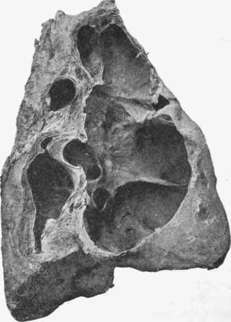
Fig. 374. - Healed cavities in lung. The figure is from a photograph of the entire lung displayed by section. There is a congeries of cavities with smooth walls and no appearance of recent action. Compare with Fig. 363.
The frequency of healing of tuberculosis of the lungs has been estimated by the author, Harris, and others, on the ground of post-mortem observation in cases which have died from non-tuberculous disease. The result is that in about 20 per cent, of persons dying from other diseases there has been at some period of life a tuberculosis of the lungs which has become obsolete.
Continue to:
My Books

