Ulcer Of The Stomach. Part 4
Description
This section is from the book "Diseases Of The Stomach", by Max Einhorn. Also available from Amazon: Diseases of the Stomach.
Ulcer Of The Stomach. Part 4
Morbid Anatomy
The peptic ulcer is found only in those regions which are exposed to the gastric juice. Aside from the stomach it is met with in the lowest part of the oesophagus and in the upper part of the duodenum. The typical gastric ulcer has a round or oval (sometimes oblong) appearance. It extends to various depths of the gastric wall, the upper part being the larger, the inferior smaller, presenting in this way more or less the shape of a funnel.
A typical ulcer looks as if it were cut out with a punch. In most instances the base of the ulcer is smooth, occasionally it is covered with tenacious greenish or brownish mucus. In microscopic sections through the margins of a recent ulcer, the ducts of the glands appear as though cut off toward the base of the ulcer. They are eaten away or digested up to the point where the tissues offer sufficient resistance to the digestive power of the gastric juice. In older ulcers, however, a reactive inflammation sets in at the periphery, leading to the formation of a callous mara, stomach: b. pylorus: c. ulcer, (From writer's observations) gin. The latter may become very much indurated, and may give on palpation the impression of a tumor, the more so if the thickened portion be situated near the pylorus. Aside from the inflammation of the narrow margin of the ulcer, the mucous membrane of the whole stomach remains in most instances normal, this being according to Rosenheim ' a principal characteristic of ulcer, which unlike cancer consists in a well-circumscribed necrotic process Laving no further influence upon the gastric mucosa. The size of the ulcer is rarely much smaller than a five-cent piece or larger than a twenty-five-cent piece, although no precise limits can be given.
Thus an ulcer not larger than a pea may exhibit all the characters of this lesion, while conversely an ulcer may gradually attain a diameter of five or six inches. Debove and Remond 1 mention a case of gastric ulcer of the size of the palm of the hand. Situation of the Ulcer. - According to Brinton,2 gastric ulcer occupies the various parts of the stomach in the following frequency: In 43 cases out of 100 the posterior surface, in 27 cases the lesser curvature, in 16 cases the pyloric extremity, in 6 cases both the anterior and posterior surfaces, often at opposite places; in 4 cases the anterior surface only, in 2 cases its greater curvature, in 2 cases the cardiac pouch. Thus about 80 ulcers in every 100 occupy the posterior surface, the lesser curvature, the pyloric sac, parts of the stomach which together form a segment of less than half of the total superficies of the organ.
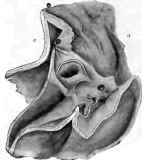
Fig.55. - Ulcer of the Stomach near the Pylorus, the latter being stenosed.
1 Th. Rosenheim: "Pathologic und Therapie der Krankheiten der Speiserohre und des Magens." Wien und Leipzig, 1891, p. 161.
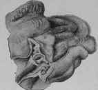
Fig. 56. The same specimen drawn in smaller proportions, in order to show the surroundings of the ulcer.
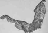
Fig. 57. - Showing the entire cross-section of an excised ulcer as it appear under the lens. The concave line forms the interior, the convex the outside of the stomach. The middle portion is deprived of the glandular layer; to the left a few glands are left. x4. (From writer's observation).
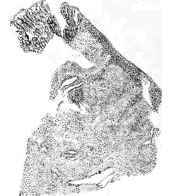
Fig. 58. - The left corner of Fig. 41, as seen under the microscope with low power.
Glands are visible to the left of the drawing, the rest consisting principally of a proliferation of cells and connective- tissue formation.
1 Debove et Rumond: "Traite des Maladies de l'Estomac."Paris p. 255. 2 W. Brinton: I. c.
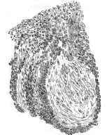
Fig. 59. - Showing One Spot of a Proliferation of Cells lying in the Centre of the Specimen (Fig. 41) resembling very much a splndle-cell aarcoma. Highly magnified.
Hence we may estimate that any part of this continued (but irregular) segment of the stomach is on an average about five times more liable to the lesion than the remaining segment formed by the cardiac sac, the anterior surface, and the greater curvature.
Nolte's1 figures do not harmonize with those just given. Nolte presents the following scale of frequency: At the greater curvature, 22; at the pylorus, 13; at the anterior wall, 3; at the posterior wall, 2; at the cardia, 1.
Welch's statistics harmonize more with Brinton's figures. Out of 793 cases collected by this eminent American writer, 288 ulcers were situated in the lesser curvature, 235 on the posterior wall, 95 at the pylorus, 96 at the anterior wall, 50 at the cardia, 29 at the fundus, 27 on the greater curvature.2
Continue to:
My Books

