The Morbid Changes In The Intestine
Description
This section is from the book "Diet In Sickness And In Health", by Mrs. Ernest Hart. Also available from Amazon: Diet in Sickness and in Health.
The Morbid Changes In The Intestine
When a person is infected with typhoid by swallowing the bacilli typhi in the water or food, the microbes make their way to the Peyer's patches and solitary glands, where they find their proper soil. They here multiply, and by their irritating presence, as well as by the poisonous substance which they excrete in the process of living, inflammation of the closed follicles is set up. The glands swell and become solid, ulceration follows, and a slough is finally separated and thrown off. The walls of the glands are succulent and vascular, and considerable haemorrhage may be caused by sloughing. The ulceration may, moreover, extend downwards through the muscular coat, even into the serous coat, and result in perforation of the intestine. If the process is gradual, the inflammation generally extends to the peritoneum; fibrinous exudations are then thrown out, and adhesions take place between the peritoneum and the thinned walls of the intestine. These inflammatory adhesions form a false wall to the intestine and prevent the escape of its contents into the abdominal cavity. The ulceration may be so severe and rapid as to cut off the blood supply of the serous coat, in which case the latter undergoes necrosis, perforation takes place, the contents of the intestine pass into the peritoneal cavity, and fatal peritonitis ensues.
Periods of the illness corresponding to morbid processes.
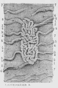
Fig. 13. - A Peyer's Patch seen from its Free or Superficial Side.
1,1,1. Afolded Peyer's Patch. 2.2. The folds which form the superficial or mucous layer of this patch. 3, 3. The grooves which separate the folds. 4, 4. Pits observed from place to place in these folds. 5, 5,5. Valvulae conniventes. 6, 6. Solitary closed follicles situated in the space between the valvulae. 7, 7, 7. Other follicles similar to the preceding but smaller. 8, 8. Closed follicles situated on the summit of the valvulae conniventes.
- Inflammation of the follicles is contemporaneous with the' first symptoms of illness. It reaches its culminating point about the tenth day. If the case is slight, resolution or absorption of the products of inflammation then takes place slowly; in severe cases the follicles ulcerate. The sloughs separate during the third week of the illness, but the process may not be completed until the fourth week. Cicatrisation of the raw surfaces of Peyer's patches begins about the end of the third week, and takes about two weeks to complete. Indiscretions of diet may, however, again set up inflammation, and Dr. Bristowe declares that the liability to perforation continues for from two to three months after the commencement of the illness.
Continue to:
My Books

