


Female Reproductive Organs
Description
This section is from the book "Instrumental Insemination Of Queen Bees", by Otto Mackensen, Kenneth W. Tucker. Also available from Amazon: Instrumental Insemination Of Queen Bees.
Female Reproductive Organs
The female reproductive organs are illustrated schematically in figure 1. Figure 2 shows a posterior exterior view of the genital opening with the sting chamber opened as in preparation for insemination.
A large part of the abdomen is occupied by two ovaries (0), each consisting of long parallel egg tubules. Each ovary leads posteriorly into a lateral oviduct (LOD) which has accordion walls permitting great expansion for temporary storage of semen in newly mated queens and of mature eggs in laying queens. The two oviducts join posteriorly to form a short passage, the median oviduct (MOD), which in turn leads into the vagina (V"). In instrumental insemination the end of the syringe must pass through the vagina and be placed in the median oviduct and the semen discharged into the two lateral oviducts.
Above the vagina lies the spermatheca (SP), a thin-walled spherical structure about 1 mm. in diameter, which is filled with a clear liquid in virgin queens. It is covered with a network of tracheae, which gives it a white appearance. It is in the spermatheca that the sperm are stored for the life of the queen. The spermathecal duct (SPD) leads from the spermatheca to the dorsal wall of the vagina.
A tonguelike structure, the valvefold (VF), projects into the vagina from its ventral wall. This structure can act as a valve to close the opening to the median oviduct when forced anteriorly against the forward wall of the vagina. It has transverse ridges which make it recognizable at times when it is exposed during instrumental insemination.
The vagina opens through the vaginal orifice (VO) into the anterior part of the sting chamber, the bursa copulatrix (BC), which is set off from the remainder of the sting chamber by a transverse fold in the ventral wall. In figure 2 this fold is stretched to form a triangle with the sting as its base. To either side of the vaginal orifice are two openings (BPO) that lead into a pair of pounches, the bursal pouches (BP). These openings can be mistaken for the vaginal orifice during instrumental insemination if the queen is not properly positioned.
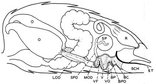
Figure 1. -Reproductive organs of the queen in approximately their natural position in the abdomen, with left side of vagina and bursa copulatrix cut away: BC, bursa copulatrix; BP, right bursal pouch; BPO, opening to bursal pouch; LOD, lateral oviduct; MOD, median oviduct; 0, ovary; R, rectum; SCH, sting chamber; SP, spermatheca; SPD, spermathecal duct; ST, sting; V, vagina; VF, valvefold; VO, vaginal orifice.
In figure 2 the sting chamber is held open as in instrumental insemination by a ventral hook (VH) placed over the last ventral plate (VP) of the abdominal exoskeleton and a sting hook (STH), which fits between the bases of the lancets of the sting (ST). Here the reproductive tract has been compressed, the bursa copulatrix opened wide to expose the vaginal orifice, and the vagina collapsed so that the valvefold lies immediately inside the vaginal orifice and occasionally is visible without any probing.
Continue to:


My Books

