Fracture Of The Sesamoid Bones
Description
This section is from the book "The Horse - Its Treatment In Health And Disease", by J. Wortley Axe. Also available from Amazon: The Horse. Its Treatment In Health And Disease.
Fracture Of The Sesamoid Bones
Fracture of the sesamoid bones is by no means of uncommon occurrence. It happens most frequently in old hunters and chasers when carrying heavy weights over deep ground, and mostly at the end of a long and tiring run.
The line of the fracture is usually transverse. Sometimes the accident is confined to one bone, but more frequently it involves both, and now and again it occurs to both fore-limbs at the same time. The bone may break through the middle, or a piece of the upper angle only may be torn away from the rest. The fracture sometimes follows upon repeated chronic sprain to the suspensory ligament, and there is reason to think that in these cases the cohesion of the bone has been diminished by an extension of the inflammatory action from the ligament to the bone to which it is attached, for it frequently occurs that prior to breakage the sesamoid bones have been for some time more or less enlarged. Slipping and false steps in making sharp turns are sometimes accountable for this injury.

Fig. 340. - Oblique and Transverse Fractures of the Os Suffraginis.
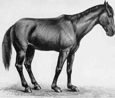
PLATE XXXIX. OSTEOPOROSIS.
Showing Disease and Descent of the Sesamoid Bones.

PLATE XXXIX. FRACTURE OF THE SESAMOID BONES.
Border Lass, aged, broke down (fractured the sesamoid bones of both fore-legs) at Ringmer Steeplechase, April 10, 1889. Photograph taken seven weeks later.
It has been said that "the accident is quite as likely to happen while the horse is at rest in his stall as under any circumstances"; but while granting the possibility of such an occurrence, we cannot subscribe to a statement for which experience affords no sort of support. We have repeatedly known horses which have been laid up for some time on account of lameness in the fetlock joint, in which perceptible enlargement of the sesamoid bones existed, to fracture their bones in the stable, or very soon after renewing work, but we have always regarded such cases as having been predisposed to fracture by a process of rarefaction of the bones arising out of inflammation extending from the sprained ligament.
Many cases of what is called "breakdown", if carefully examined after death, would be found to result from a giving way of the bones in this weakened condition, and the removal of a fragment by the partially-separated ligament (Plate XXXIX).
These cases are attended with lameness more or less severe, but in the slighter accidents there may be but little distortion of the fetlock joint, and the writer has found that in course of reparation the ligament at its point of attachment with the sesamoid bone becomes ossified.
Symptoms
Fracture of the sesamoids results in sudden lameness, but in degree varying with the nature of the fracture. When this is completely across the bone, the broken fragments are forced asunder by the weight imposed upon them, and the fetlock joint, having lost the support of the suspensory ligament, sinks towards the ground. The weight, now falling more directly upon the heel, gives the toe an inclination upwards. This deformity may not occur immediately, and sometimes only appears two or three days after the accident, at which time there is much swelling, heat, and pain in the part. It may be that only one sesamoid bone may be fractured, in which case the fetlock joint inclines-inward or outward, according as the one side or the other is affected. Although the broken pieces may be much displaced, crepitus may sometimes be induced by fixing the suspensory ligament above and forcibly flexing the fetlock joint.
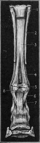
Fig. 341. - Fracture of the Sesamoid Bones.
1, Carpal tendon cut away to show suspensory ligament. 2, Small metacarpal bone. 3, Suspensory ligament. 4, Branch of same. 5, Fractured sesamoid bones. 6, Deep sesamoid ligament. 7, Superficial sesamoid ligament. 8, Intersesamoid ligament.
Treatment
Very little can be done in these cases to fit the horse for remunerative work, but when it is required to put it to the stud, in some cases it may be made serviceable for the purpose.
The strain should be taken off the suspensory ligament by the application of a high-heeled shoe. The joint should then be supported by a starch bandage, carried from the coronet upwards to the middle of the canon. This having been clone, the animal should be placed in slings and kept there eight or ten weeks, or longer if necessary, as quiet as possible.
In complete fracture, the fetlock joint is sure to remain deformed and enlarged to a greater or less extent. A repetition of blisters after the patient has been taken out of the slings will help to reduce the enlargement, and give further tone to the injured parts.
Continue to:
My Books

