Parasites Of The Digestive System Of The Horse. Continued
Description
This section is from the book "The Horse - Its Treatment In Health And Disease", by J. Wortley Axe. Also available from Amazon: The Horse. Its Treatment In Health And Disease.
Parasites Of The Digestive System Of The Horse. Continued
Two other nematode worms are found in the intestine of the horse. Both of them deposit their eggs beneath the mucous membrane, giving rise to small tumours. The two parasites are known as (1) the Strongylus armatus and (2) the Strongylus tetracanthus. The latter is the most common and the most destructive. The Strongylus armatus is distinguished by its large mouth, which is armed with a row of cutting teeth arranged close together. The Strongylus tetracanthus has, in addition to the row of teeth like the Strongylus armatus, four large spines, from which the name is derived, and also inside the mouth a row of sharp hooks. It will be evident, therefore, that the creature is well provided with offensive weapons. The worm is constantly found accumulated in the large bowel, frequently in company with the Strongylus armatus.
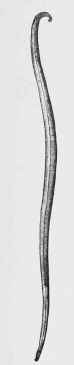
Fig. 274. Ascaris (about 1/2 nat. size).
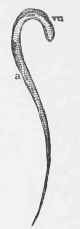
Fig. 275. - Oxyurus of Horse - Female (natural size).
vu, Vulva. a, Anus.
In young animals a serious mortality is often caused by the invasion of this worm in large numbers, as both the parent worm and the young ones are true blood-suckers, and the embryos begin their ravages as soon as they escape from the cysts in which they are coiled up under the mucous membrane, in the manner seen in the illustration below, which is taken from a portion of the caecum.
In the case of these two parasites, medical treatment has not hitherto proved very successful. Turpentine, chinosol, perchloricle of iron are the most promising remedies. Colts, the animals which suffer most from the invasion of the parasite, may receive san-tonine in doses of 10 grains in a ball, or mixed with the food every day for three or four days, to be followed by a dose of linseed-oil.
Other nematodes have been described by writers, but they are not of very frequent occurrence, and it does not appear that they have been found among horses in this country. Information regarding them may be found in Neumann 0n Parasites, from which work some of the illustrations of the present chapter are reproduced.
Very few of the parasites of the next class, cystic worms or Hat-worms (Hat helminths), inhabit the intestines of the horse.
The common name tape-worm is given to these parasites. In the horse the few tapeworms which infest the intestines are remarkable for their small size in comparison with other varieties which are found in cattle, sheep, and dogs.
The three varieties are: the Taenia per-foliata, which is something under 2 inches long and 3/8 inch in width; Taenia plicata, about 3 1/2 inches long and 3/8 inch in width; and Taenia mamillana, which is about an inch in length, and a little more than 1/8 inch in width. (Fig. 276.)
The perfoliata, which is most common, is distinguished by the presence of a peculiar appendage, forming a kind of collar, round the neck. The parasite inhabits the caecum, seldom being seen in other parts of the intestinal canal. (Fig. 277.)
Taenia plicata is said to be found in the small intestine; sometimes in the stomach (fig. 278). The Taenia mamillana (fig. 279) is also said to exist in the small intestine, but there is no record of its having been found among horses in this country. All these tape-worms are peculiar in being unarmed, that is, are not provided with a double row of hooks, which are common in other varieties. Nothing is known of the hydatid stage, which forms the intermediate condition between the tape-worm embryo and the mature parasite.
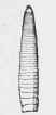
Taenia mamillana (nat. size).

Taenia perfoliata (nat, size).

Taenia plicata (nat. size).
Fig. 270. - Tape-worms.

Fig. 277. - Teania perfoliata, Cephalic Extremity (enlarged 7 diameters).

Fig. 278. - Taenia plicata, Cephalic Extremity (enlarged 7 diameters).

Fig. 279. - Taenia mamillana, Cephalic Extremity (enlarged 15 diameters).
There are no indications whatever of the existence of the worms during the life of the horse which they infest, and consequently no treatment has ever been attempted. A remarkable case is recorded of the existence of nearly all the parasites which have been described in one horse which was examined by Veterinary Surgeon Krause. There were found 519 Ascaris megalocephala, 191 Oxyurus curvula, 214 Strongylvs armatus, many thousands of Strongylus tetracanthus, 69 Taenia perfoliata, 287 Filaria papillosa, and 6 Cysticercus fas-ciolaris.
To complete the history of the parasites which infest the digestive organs of the horse it is necessary to allude to some which are found occasionally in the liver. The fluke (Distoma hepaticum) (fig. 280) sometimes effects an entrance into the liver ducts of horses, especially colts, which are feeding on wet pastures where the embryos and larval forms of the parasite are abundant. Sheep, as is well known, are destroyed in thousands in some localities by the invasion of this parasite, which causes the disease known as rot. A few cases are recorded of foals and colts having suffered from the accidental invasion of the fluke, but the disease among horses must be looked upon as entirely exceptional. The fluke is not one of the worms which finds a host in that animal under ordinary circumstances.


Fig. 280. - Liver Fluke (Distoma hepaticum, Linn).
A, Showing Anatomical details. b, Natural size. C, Ciliated Embryo, or Young Distome.
Another parasite which is found in the liver of the horse is the cystic stage of an extremely small tape-worm found in the intestines of the dog, the Taenia echinococcus. The worm, when fully grown, is on an average about 1/4 inch in length, and never exceeds 1/2 inch, but in its cystic (hydatid) stage it is one of the largest which exists.
The Cysticercus echinococcus is found frequently in the liver, and occasionally the lungs, of cattle and sheep; the cysts varying in size from that of a grape to that of an orange, as a rule, but now and then they are found of an enormous bulk. Each cyst contains a fluid in which are found floating a number of tape-worm heads, myriads of which are observed growing on the interior of the cyst. In one form of the parasite small cysts, or daughter-vesicles as they are called, are found abundantly in the fluid. This peculiarity has given rise to a division of the parasite into two classes: -
1. The Echinococcus altricipariens, in which the secondary vesicles exist.
2. The Echinococcus scolicipariens, in which they are replaced by the small spots on the membrane, and in the fluid the tape-worm heads (Scolices).
The presence of these hydatids in the liver and other organs of animals is often not attended with any indications of disease, even when the liver is so filled with the cysts as apparently to replace the normal structure.
On the serous membrane of the chest and abdomen small wandering echinococcus cysts are occasionally found. There is also a nematode worm (Filaria) which has been found in the peritoneal and pleural cavities of the horse, ass, and mule. It does not appear to have been recorded, however, among the parasites of the horse in this country.
In the circulatory system of the horse, parasites are occasionally encountered, as the Surra parasite, found in the blood of horses in India, and the embryos of the Strongylus armatus and Strongylus tetracanthus, which locate themselves in the anterior mesenteric artery, and cause a well-marked aneurism. It is comparatively common in the ass. Parasites in the nerve-centres, or in the organs of special sense of the horse, are extremely rare. There is one case recorded by Woodger of the presence of a hydatid in the brain of a horse. In this case the animal suffered from the same kind of giddiness and tendency to turn in one direction as is known to be characteristic of a sheep similarly affected with hydatid in the brain, and there are a few cases reported of the discovery of the embryos of the armed Strongylus in the blood-vessels of the brain.
Cases have also been reported of the presence of hots (larvae of the (OEstrus equi) in the brain cavity of the horse, and also in the spinal canal of a pony.
Among the organs of special sense, the eye of the horse seems to be the only one which is invaded by parasites. It is recorded that Van Setten removed a pentastome from the right eye of a horse, and in horses in India the presence of a nematode worm is extremely common. The parasite is easily removed by puncturing the cornea and allowing the aqueous humour to escape, carrying with it the worm.
A minute worm (Filaria palpebratus) is occasionally found under the eyelids of the horse, causing irritation, with swelling of the eyelids and an abundant secretion of tears.
Continue to:
My Books

