The Crucial Ligaments
Description
This section is from the book "The Horse - Its Treatment In Health And Disease", by J. Wortley Axe. Also available from Amazon: The Horse. Its Treatment In Health And Disease.
The Crucial Ligaments
These are two thick short bands situated in the notch which separates the two condyles, where they cross each other like the lines of the letter X. The anterior branch is attached to the spine on the upper end of the tibia, and to the intercondyloid notch and the external condyle. The posterior branch is united by its lower extremity to the superior and posterior part of the tibia, and by its upper end to the notch between the condyles and to the internal condyle. The Posterior Ligament is practically the posterior section of the capsular ligament of the femoro-tibial articulation. It is attached to the femur, behind and above the condyles, and to the posterior part of the head of the tibia, just below its articular margin. It joins the lateral ligaments, uniting the femur and tibia on either side, and its inner face is lined by synovial membrane.
The Interarticular Fibro-cartilages are the crescentic pieces of dense fibro-cartilage upon which the condyles of the femur are made to rest on the head of the tibia or second thigh. They are hollowed out above for the reception of the condyles, for which they form a bed. The outer cartilage is attached in front to the base of the spine on the head of the tibia, and behind by two slips, one to the notch between the condyles, and the other to the upper and posterior part of the tibia.
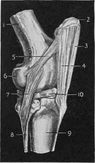
Fig. 359. - Femoro-tihial Articulation or Stifle-joint.
1, Femur. 2, Patella. 3, Middle straight ligament. 4, External straight ligament. 5, External lateral ligament of patella. 6, Outer condyle. 7, External lateral femoro-tibial ligament. 8, Fibula. 9, Tibia. 10, External inter-articular cartilage.
The inner cartilage is attached in front and behind to the base of the spine on the head of the tibia.
This joint possesses three synovial membranes, one of considerable extent enclosing the articular surfaces of the patella, and the two ridges or trochlea in front of the femur, and one to each condyle of the femur and its corresponding half of the articular face of the tibia.
The movements of this joint are essentially those of flexion and extension, but it also enjoys a limited power of rotation.
Continue to:
My Books

