Zoological Distribution
Description
This section is from the book "The Horse - Its Treatment In Health And Disease", by J. Wortley Axe. Also available from Amazon: The Horse. Its Treatment In Health And Disease.
Zoological Distribution
Although solipeds would appear to be the only animals affected by it, it has been said to have occurred in cattle, and in referring to this Pallin remarks: "Care should be taken not to confound it with a disease found among the cattle in Guadeloupe known under the name farcin de boeuf, and due to a bacillus discovered by Nocard, and which Metschnikoff describes as a streptothrix. This disease is only transmissible to cattle, sheep, and guinea-pigs, but does not affect horses or donkeys."

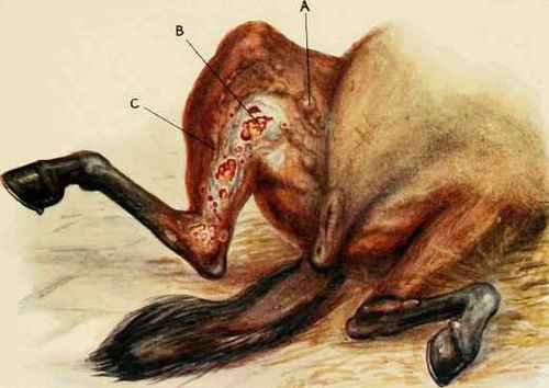
I. AA, abscesses in the course of formation; B, ulceration of skin and subcutaneous tissue following upon breakage of abscesses: c, inflamed lymphatic vessels. 2. A. abscess in the course of formation; B, ulceration of skin and subcutaneous tissue following upon breakage of abscesses; c, inflamed lymphatic vessels.
Diagnosis
Contagious lymphangitis until comparatively recent times and in every country where it is known has been confounded with farcy. It is only since the adoption of bacteriological methods, and later by reason of the mallein test, that any difference has been recognized. Examination of the matter taken from abscesses in the skin shows at once the crypto-coccus as a minute lemon-shaped organism having a highly refractile double outline. Moreover, the mallein test fails to give the fever reaction and the local swelling commonly associated with glanders.
Mallein when used alone leaves the mind in doubt, but if the organism be present, then there need be no reason to hesitate to give an opinion.
Points of difference between epizootic lymphangitis and farcy are at first sight not great, but in the matter of diagnosis nothing short of finding the cryptococcus on which the disease depends should be considered sufficient, and especially as this is by no means difficult of accomplishment.
The time which elapses between the reception of the poison and the outbreak of disease is, like glanders, very variable, and may be expected any time between five weeks and four or five months, and in some instances it has been known to exceed a year.
One attack of the disease does not prevent a second.
Under the most favoured circumstances the average mortality is said to be about " ten to fifteen "per cent. but in a disease which lasts so long, is so liable to recur after long periods, and is generally so uncertain and unsatisfactory, it would be difficult to say exactly what the mortality may be, and in large studs of horses to trifle with a disease of this description would be carrying risk to the verge of ruin.
Symptoms
It is strange that a disease which is considered sufficiently serious to be scheduled as a contagious and dangerous malady should have practically no constitutional symptoms. The importance of the disease evidently does not dwell in its life-destroying danger, but in the fact of its contagious and crippling nature.
The value of a horse depends entirely on our power to use him, and in this disease he is for the most part and for long periods removed from our will to do so. Moreover, so long as he remains in our stables he is a source of danger to others, and although he may ultimately become well again he is nevertheless likely to infect his companions while doing so, and above all to leave the stable a centre of infection.
We were surprised a short time ago in looking over an infected stud to find all the horses, notwithstanding the disease, in good condition, full of flesh, feeding well, the temperature and pulse normal, and the coat sleek.
The local symptoms invariably attack the skin, sometimes the mucous membranes of the nose and eye are also involved, especially as the result of auto-inoculation while scratching or rubbing infected places.
All parts of the body, wherever there is a wound to admit the virus, are liable to become the seat of the disease.
From four to six weeks after infection a small hard nodule appears on the site of inoculation, and the lymphatics about it may be felt beneath the skin, or the latter may be first enlarged or "corded " to the feel.
In some cases it has been noticed that a general enlargement of the limb occurs, and the lymphatic enlargement is only recognized when the swelling has subsided.
The primary nodule and lymphatic vessels continue to enlarge until the latter stand out as distinct lines radiating from the former. At first the nodule is hard and small, but ultimately reaches the size of a walnut or a hen's egg. At this time it is soft and fluctuating, and soon breaks and discharges a quantity of pus, which is remarkable for its thick sticky nature, a character which does not belong to the pus of farcy. This is soon followed by the development of granulations, which not only fill up the cavity but likewise extend beyond it in the form of proud-flesh. With the discharge of pus the edges of the wound have a tendency to fall in, but this is soon prevented, either by the development of granulation and the filling up of the wound, or by the ulceration and loss of the skin.
In all cases the wound has an indurated base, the surrounding connective tissue having taken on a slowly progressive inflammation, of which abscess is the result.
In some instances there are chains of abscesses running along the line of the thickened lymphatic vessels, and breaking out at varying intervals of time, so that abscesses entire, broken, and in process of repair may sometimes be seen side by side.
When a swelling is cut into in the early period of the disease, it is found to be hard and grayish-white in colour. There is a considerable connective tissue new growth in the part, and as the point of ripening is reached a dark vascular area appears, in the midst of which suppuration proceeds and continues until an abscess is developed.
Continue to:
My Books

