Cocoa Microscopical Examination
Description
This section is from the book "Tea, Coffee, And Cocoa Preparations", by Guilford Lawson Spencer . Also available from Amazon: Tea, coffee, and cocoa preparations.
Cocoa Microscopical Examination
For a thorough study of cocoa preparations, a microscopical examination is indispensable. An accurate knowledge of the structure of the cocoa bean and of the substances used as adulterants is necessary for the successful carrying out of this investigation. While this information is only to be gained by actual study of the materials in question, its acquirement is greatly facilitated by the use of descriptions and illustrations.
The literature of the subject will be found somewhat contradictory (even with comparatively recent writers) in some details, but nothing of importance in investigations for detection of adulteration seems to be subject for debate at the present time. The works of Moeller and Mace will be found to furnish valuable assistance in investigations of this kind. The microscopical characteristics of the starches and other materials used for adulteration have been so well described in various works and in previous bulletins of this Department that any detailed description of them seems unnecessary here.
1 Mace, Los substances alimentaires etudiees au microscope. 2Chem.News, 62, 99.
The cocoa bean is inclosed in a thin, brittle, reddish brown seed coat, called the husk or shell. On the surface of the husk are often Found numerous delicate, tubular cells, which come from the pulp of the fruit. The important structures of the husk proper are the following:
(1) The epidermal layer.
(2) The loose parenchyma.
(3) The fiber bundles with small spiral cells.
(4) The layer of characteristic thick-walled cells.
After softening the husk by soaking the bean in water, a portion of the epidermal layer is readily torn away with the forceps, freed from adhering fragments of the adjacent tissue, and placed on the slide for examination. It is found to consist of a layer of moderately thick-walled, somewhat elongated, irregularly polygonal cells (see Plate XLVI). By careful dissection and careful manipulation of the light and the micrometer screw, a layer of exceedingly delicate, transversely elongated cells can be seen to lie directly under the layer just described, but it is so very delicate that it is rarely seen in the examinations of preparations of cocoa for adulterants, and is consequently of almost no importance in such investigations. If some of the underlying tissue exposed by the removal of the epidermis be transferred to a slide and dissected apart, it will appear as a mass of loosely aggregated, rather large, thin-walled, slightly elongated cells, those constituting the inner layers containing a large amount of mucilaginous matter that swells up in contact with water and ruptures them. This parenchymatous tissue, which makes up the greater part of the husk, is pierced in all directions by small, ramifying fiber bundles inclosing small spiral cells and stone cells; near the inner surface of the husk it is interrupted by a single layer of small, very thick-walled cells (see Plate XLVI), which are very characteristic and withstand the disintegrating processes of manufacture better than any other part of the husk. In fact, these processes are often carried so far that it is only by very diligent search that one is able to find any recognizable structures besides these cells and the starch grains of the cotyledons. For the detection of the presence of husks in cocoa preparations, these thick-walled cells are first sought for; and after these the epidermal and parenchymal structures. The fiber bundles, with inclosed spiral cells, are not readily distinguished from those of the cotyledon.
Bull.NO.I3 Div.Of Chemistry. Plate XLVI
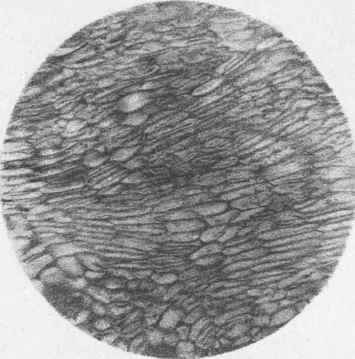
Cocoa X75
Epidermis of Husk.
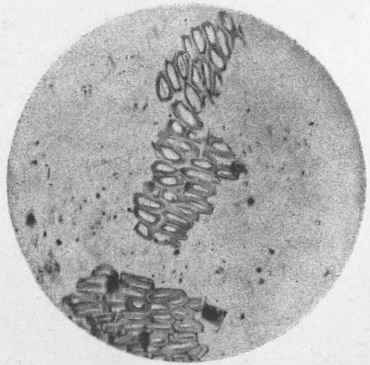
Cocoa Husk Xll5
Characteristic thick-walled cells.
A.Hoen & Co.Heliocaustic.Baltimore.
If the brown husk be entirely removed from the remaining part of the bean, a thin, transparent membrane will be observed, which partly comes away with the husk and partly remains adherent to the bean proper, dipping into all the clefts and plications of the latter. When a portion of this membrane is examined with the microscope, it appears as a single layer of small polygonal cells which are filled with granular matter (see Plate XLVII). By careful manipulation one or more layers of parenchymatous cells can be found underneath the layer just described. Adherent to this membrane, especially to the folds entering the clefts of the cotyledons, are numerous yellow, club-shaped masses
Bull.NO.I3 Div.Of Chemistry. Plate XLVII
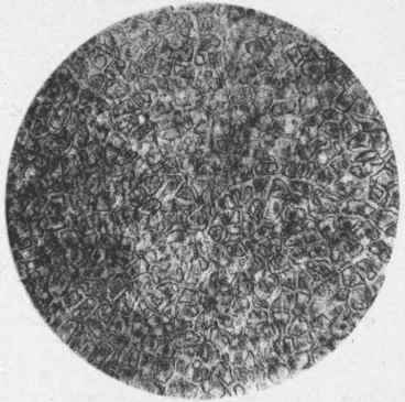
Cocoa Husk Xll5
Thin inner membrane.

Cocoa Xll5 Section Of Cotyledon.
A.Hoen & Co.Heliocaustic,Baltimore.
20393 - No. 13 - 6 of cells, known as "Mitscherlich bodies." They are now considered to be epidermal hairs, but it is not decided as to whether they belong to the membrane just described or to the surface of the cotyledon. Neither the settlement of this question nor the membrane and hairs, are of any great importance for our purpose, since both of these structures are very rarely met in recognizable form in the commercial preparations.
After the removal of the husk and the membrane just described, the two fleshy, much-folded cotyledons, or seed leaves, remain, inclosing the radicle (the embryo stem of the undeveloped plant) at the larger end of the bean in a manner not a little suggestive of the retracted head of a turtle. Examination of a thin section shows the cotyledon to be made up of comparatively thin-walled, closely packed, polygonal cells (see Plate XLVII). The most of these cells are filled with starch grains, fat, and albuminous material; isolated or small groups of cells are homogeneously filled with the reddish brown to violet pigment, cocoa red. Fiber bundles with spiral cells, similar to those of the husk, occur in the veins.
Continue to:
My Books

