Lumbar And Sacral Plexus And Nerves Of Lower Limb. Continued
Description
This section is from the book "Handbook Of Anatomy For Students Of Massage", by Margaret E. Bjorkegren. Also available from Amazon: Handbook Of Anatomy For Students Of Massage.
Lumbar And Sacral Plexus And Nerves Of Lower Limb. Continued
Obturator nerve arises from the second, third, and fourth lumbar nerves. The nerve emerges from the inner border of psoas behind the common iliac vessels. It passes forwards with the obturator artery, and goes through the groove in the thyroid foramen, where it divides into two branches-superficial and deep. This nerve supplies the muscles and skin on the inner side of the thigh.
The Superficial part of the obturator nerve enters the thigh beneath pectineus, and, passing down the inner border of adductor longus, anterior to gracilis, it finally divides into two terminal branches, one of which enters Hunter's canal.
Branches
Muscular to adductor longus, gracilis, adductor brevis, and pectineus (occasionally).
Cutaneous becomes superficial in the middle third of the thigh, and supplies skin of the lower two-thirds of the inner side of the thigh, and ends in the subsartorial plexus. The terminal branch, which enters Hunter's canal, ramifies over the femoral artery.
The Deep part of the obturator nerve pierces obturator ex-ternus and passes down between adductor brevis and adductor magnus; it then passes through adductor magnus, and, entering the popliteal space, terminates by supplying the knee-joint.
Branches
Muscular to obturator externus, adductor magnus and adductor brevis (if not already supplied by the superficial part).
Articular to the knee-joint.
The Anterior Crural nerve arises from the second, third, and fourth lumbar nerves behind the obturator nerve. It is formed in the substance of psoas, and, emerging from its outer border, it passes down between psoas and iliacus, and enters the thigh by passing under Poupart's ligament on the outer side of the femoral vessels. In Scarpa's triangle it breaks up into branches to supply the front of the thigh.
Branches
In the abdomen: Muscular to iliacus.
In Scarpa's triangle: Muscular to pectineus, sartorius, and quadriceps extensor.
Articular to the hip and knee joints.
Cutaneous
The cutaneous branches are in three sets-middle and internal cutaneous and long saphenous.

Fig. 70. - Muscles and Cutaneous Nerves of Leg (Posterior View).
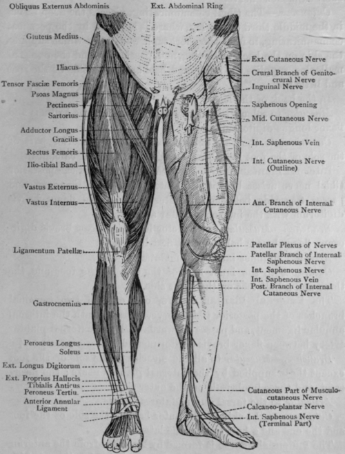
Fig. 71 - Muscles and Cutaneous Nerves of Leg (Anterior View).
Middle Cutaneous nerve arises in two parts-the external and internal. They supply the skin on the lower three-fourths of the front of the thigh, and end in the patellar plexus.
Internal Cutaneous nerve lies in Scarpa's triangle on the outer side of the femoral vessels, over which it crosses, and, dividing into three branches, all of which terminate in the patellar plexus, supplies the skin on the lower two-thirds of the inner side of the thigh.
The Long Saphenous nerve arises in Scarpa's triangle. It passes down with the femoral vessels through Hunter's canal, at the lower end of which it crosses over the tendon of adductor magnus and becomes cutaneous on the inner side of the knee-joint by passing between sartorius and gracilis. It passes down the inner side of the leg with the internal saphenous vein, and supplies the skin of the front and inner side of the leg and posterior half of the dorsum and inner side of the foot.
The Patellar Plexus is formed by the branches of the cutaneous nerves supplying the skin in front of the knee-viz.,
Continue to:
My Books

