2. Haemorrhage From The Nutrient Arteries
Description
This section is from the book "A Manual Of Pathology", by Joseph Coats, Lewis K. Sutherland. Also available from Amazon: A Manual Of Pathology.
2. Haemorrhage From The Nutrient Arteries
As these vessels run in the substance of the brain the haemorrhage is always cerebral and rarely extends to the meninges. It might be supposed that as the nutrient vessels are small the haemorrhage from them would be small, and in many cases it is so; but when bleeding has once begun, the blood tearing the brain substance ruptures other vessels, and there is often a considerable effusion of blood.
Haemorrhage of any consequence rarely occurs from the cortical nutrient vessels, but is very common from the central nutrient arteries.
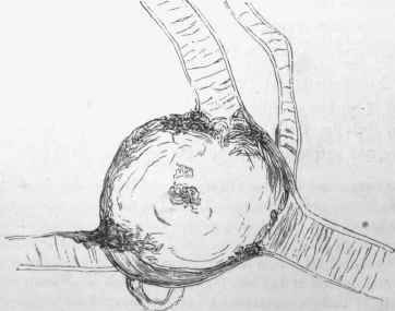
Fig. 328. - Sacculated miliary aneurysm of a nutrient artery of the brain. The aneurysm is about the twentieth of an inch in diameter, x 27.
The circumstances of these latter go far to explain this. They arise from large stems, mainly from the middle cerebral immediately after its origin from the internal carotid. It is clear that the blood here will be at a pressure not much less than that of the aorta, and any variations of pressure will tell readily. On the other hand, the cortical vessels mostly arise from fine vessels in which the blood-pressure has been reduced by successive division and sub division.
As to the Causes of haemorrhage in these arteries, Aneurysm again plays the most important part. As the arteries are small so are the aneurysms, but they are numerous in the same person. Such aneurysms have been called by Bouchard and Charcot Miliary aneurysms. They occur in every region of the brain, but are most readily detected on the surface of the convolutions, where, on stripping off the pia mater from the convolutions, they may be seen as small red or brown spots. When examined under the microscope they have all the characters of ordinary aneurysms. Most of them are sacculated (Fig. 328), but some are fusiform (Fig. 329). It is stated that the cause of their formation is a sclerosis of the walls of the arteries, involving first a formation of round cells in the external coat with subsequent development into fibrous tissue. According to Recklinghausen, however, the first lesion is a rupture of the media - an origin which, considering the mode of formation of aneurysms generally, seems a very probable one. In that case the sclerosis of the wall is to be regarded as the result of an inflammation secondary to the injury. Miliary aneurysms are mostly met with in old people; in persons above fifty cerebral haemorrhage is, in the larger proportion of cases, due to rupture of miliary aneurysms.
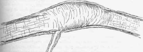
Fig. 329. - Fusiform miliary aneurysm, x 27.
In a case observed by the author there was frequently in the arteries a fatty degeneration affecting chiefly the internal coat. This was present in patches in a large numbef of vessels of small size. In connection with it there was sometimes a partial dilatation, an aneurysm obviously forming, and also fully formed aneurysms. Older and more recent haemorrhages were connected with the aneurysms, and there was one very large fatal haemorrhage.
Although the aneurysms are present in all regions of the brain, rupture seldom occurs except in those of the central arteries. The explanation of this has already been indicated, and it has been mentioned that the lenticulo-striate branch is pre-eminently that from which haemorrhage occurs.
Atheroma, with increased blood-pressure, is occasionally a cause of haemorrhage from the nutrient arteries. It is difficult to understand how atheroma, which consists in a thickening of the intima, should lead to haemorrhage. It has been pointed out, however (see p. 484), that the atheromatous patch often produces injury and rupture of the media, thereby leading to aneurysm in some cases. In like manner, in such small thin-walled vessels as the nutrient arteries it may predispose to rupture. It is doubtful if rupture actually occurs without an increase in blood-pressure. Hence the coincidence of atheroma with chronic Bright's disease - in which the general blood-pressure is raised - not infrequently leads to cerebral haemorrhage.
Atheroma is not a frequent disease in. the smaller arteries of the body. It is very common in the larger arteries of the brain, and extends even to the smaller branches of these arteries in the sulci. It does not usually affect the cortical nutrient arteries, but not infrequently extends to the larger central arteries, especially those going to the basal ganglia. As these arteries are, for their size, exposed to a higher blood-pressure than others, and as they are surrounded by the soft brain substance, they rupture more readily. It will be noted that it is these same arteries, and more particularly the lenticulo-striate branch, which are most frequently the seat of rupture from aneurysm.
Continue to:
My Books

