


4. Pulmonary Haemorrhage
Description
This section is from the book "A Manual Of Pathology", by Joseph Coats, Lewis K. Sutherland. Also available from Amazon: A Manual Of Pathology.
4. Pulmonary Haemorrhage
Haemorrhage occurs in the lungs under a considerable variety of circumstances and presents many different appearances.
The Haemorrhagic infarction is the form most frequently met with after death. It results from obstruction of the pulmonary artery, and this is in the great majority of cases due to embolism, but in a few may be the result of thrombosis. In cases of extreme passive hyperaemia, there may be such stagnation of the blood in the pulmonary artery in some parts as to induce coagulation, but this is very rare.
The pulmonary artery in itself and its branches is an end-artery, but various local circumstances frequently interfere with the formation of the infarction. The capillaries of the lung are wide, and may act in the same way as anastomosing vessels so as to keep up the circulation and prevent engorgement. In addition, the bronchial artery nourishes the lung tissue and even forms communications with the pulmonary artery. The infarction is least likely to occur where the affected piece of lung tissue is completely surrounded by lung tissue whose capillaries communicate. The existence of the pleura at one or several surfaces will interfere with such communication, and hence the infarction is more common at edges, where two or three surfaces are covered with pleura, than in the substance of the lung or at its lateral and posterior aspects.
The observation of Pitt confirms these statements in so far as that blocking of the pulmonary artery is frequent without the occurrence of the ha>morrhagic infarction. This author asserts, in opposition to Cohnheim, that thrombosis of the pulmonary artery is a frequent lesion.
The haemorrhagic infarction presents itself as a limited piece of condensed lung tissue, which may be often recognized by its dark colour seen through the pleura, but is more easily discovered by handling the lung, when the solid mass is readily detected. It is more or less wedge-shaped, the base being at the surface. It is usually peripheral and most frequently at an edge of the lung. It presents great varieties in size up to nearly half the lung, but it is most commonly of moderate dimensions. When recent, the infarction looks on section like a recent blood clot, and has almost a similar smooth surface, from which circumstances the term Pulmonary apoplexy used to be given to the condition from the analogy of haemorrhage in the brain, which produces cerebral apoplexy. As time goes on the colour merges into brown, and the appearance may come to resemble that of red hepatization. As blood fills the air spaces the piece of lung is more bulky than that surrounding it. The infarction, therefore, is often seen as a rounded bulging beneath the pleura, or, when the lung is divided it stands at a higher level than the surrounding tissue. The pleura over the infarction is usually covered with a layer of fibrinous exudation of a yellow colour, and the pleural cavity contains fluid, often in considerable quantity.
Search should always be made for the obstruction in the artery. The arteries in the infarction itself may be filled with coagulum, but the plugging generally extends beyond its apex, the actual embolus being usually some little distance on the proximal side of the infarction. An endeavour should also be made to find the source of the embolus.
Under the microscope, the affected parts of the lung show, as in Fig. 358, an enormous aggregation of red corpuscles in the lung alveoli and finer bronchial tubes, with distension of the pulmonary capillaries. The absolute filling out of the alveoli with blood to the entire exclusion of air indicates that there has been a leakage from the capillaries gradually filling up the alveoli and expelling the air. There is usually hardly a trace of fibrine to be seen, merely red corpuscles which have escaped by diapedesis, and some catarrhal cells. Such cells are nearly always present, the blood irritating the epithelium and including their production.
If the patient live for some time after the occurrence of the infarction certain changes occur in it, but there are few actual observations bearing on this point. A case observed post mortem by the author gave definite indications as to the course of events. The symptoms during life indicated the formation of an infarction months before death. The conditions observed were those of partial recovery from the lesion. The pulmonary tissue was brown and partly condensed in a limited area. The artery supplying this area showed a plug consisting of connective tissue formed by the process of organization described at p. 101. The circulation had been partly restored by this process. It is apparent from this observation that in some cases the infarction clears away; the blood is discharged by expectoration, and the circulation is re-established, but the portion of lung is unduly pigmented. In other cases the portion of lung gradually shrinks, and the ultimate result is a pigmented induration. Indurations, probably of this origin, are not infrequently met with in cases of valvular disease. In other cases, again, the portion of lung tissue dies and sloughs, so that a cavity forms. Sometimes the slough decomposes, and we have all the features of gangrene of the lungs. It may seem strange that necrosis, which is the regular result in other organs, is not of more constant occurrence in the lungs. It is to be remembered, however, that the lung tissue in the midst of the infarction is still nourished by the bronchial artery, and that, while the capillaries and actual walls of the alveoli may die, the interlobular connective tissue may survive, and may even use the necrosed tissue as pabulum.
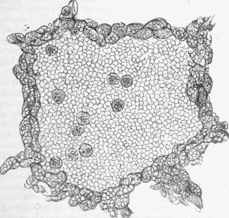
Fig. 358. - From a haemorrhagic infarction of the lung. The alveolus is filled with red blood-corpuscles, with one or two large catarrhal cells. In the wall of the alveolus the capillaries are greatly distended with blood, x 350.
Conditions resembling in general and microscopic appearances the haemorrhagic infarction occur in extreme cases of passive hyperemia. This is not surprising as the infarction is itself the result of stagnation of blood. But the haemorrhagic condensations produced by passive hyperemia have not the locality or limitation of the proper infarction.
There are also haemorrhages in scurvy, hemophilia, haemorrhagic small-pox, etc. haemorrhage is very frequent in phthisis pulmonalis, as will be subsequently described. It is asserted also that there may be rupture of a branch of the pulmonary artery from fatty degeneration of its walls, as we may have haemorrhage in the brain from atheroma., but this is excessively rare.
Haemorrhage is not uncommon from Tearing or Rupture of the lung, by a penetrating instrument or a broken rib. The blood in this case will partly escape into the pleura, but it will also collect in the cavity torn in the lung and infiltrate neighbouring parts of the parenchyma. According to Rokitansky blood thus effused may be encap-suled and subsequently infiltrated with lime salts like a foreign body.
When the haemorrhage is from the bronchial mucous membrane or the lung alveoli, then it appears in the sputum. To some extent, however, the blood remains in the air passages, and if it be in considerable quantity it may even be carried by Insufflation into the lung alveoli. The blood, in this case is mixed with air, and there is no such condensation as that which occurs in the haemorrhagic infarction. In the lung alveoli the blood acting as a foreign body irritates the tissue and a catarrhal inflammation may result. In such cases large catarrhal cells occupy the alveoli abundantly wherever the blood has penetrated, and these cells may be deeply stained with the colouring matter of the blood.
Literature
Rokitansky, Lehrb., iii.; Zenker, Beitr. zur norm. u. path. Anat. d. Lungen, 1862; Hertz, in Ziemssen's Handb., v.; Cohnheim, Allg. Path., 1882, i., 501; Gerhardt, Volkmann's lectures (Syd. Soc. transl.), 1876, 2nd ser., p. 261; Pitt, Path, trans., xliv., 1893; Fujinami, Virch. Arch., clii., 1898; Scriba, (Fat Embolism) Untersuch. u. die Fettembolie, Deut. Zeitsch. f. Chururgie, 1879, B. xii., § 110 (literature).
Continue to:


My Books

