6. Chronic Rheumatic Arthritis
Description
This section is from the book "A Manual Of Pathology", by Joseph Coats, Lewis K. Sutherland. Also available from Amazon: A Manual Of Pathology.
6. Chronic Rheumatic Arthritis
As a rule this disease affects many joints. The condition is sometimes designated Rheumatic gout. As the changes are virtually the same as in Arthritis deformans, and the two conditions run into one another, we may describe them together. In arthritis deformans the disease is partial, being confined perhaps to a single joint, and the lesions attain a much greater development than in general articular rheumatism. It is also much more a disease of old people, and is met with chiefly among the poor. According to Lane the conditions to be here described are not rheumatic in their origin, but are the result of undue pressure and friction on the ends of the bones. To this he ascribes the frequency of their occurrence amongst working people. This view is probably a partial one, but the cause suggested may be regarded as a contributory element.
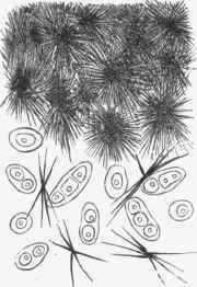
Fig. 295. - Cartilage of joint in gout, with crystals of urate of sodium. The salt is in stellate crystals which are nearly continuous at upper part of figure; which corresponds to surface of joint. x 200. (Cornil and Ran-vier).
The inflammatory phenomena appear first in the synovial membrane and the cartilages. The synovial fringes enlarge by a slow process of inflammation, and the villous projections increase in number and become more prominent. Not uncommonly pieces of cartilage develop in the fringes, originating in the cartilage cells which exist normally there, and these pieces of cartilage, being usually pedunculated, act very much like free bodies in the joint. Portions of the prominent outgrowths may get actually separated, and so we may have loose bodies in the joints. This cartilage also sometimes undergoes ossification in whole or in part. In the early stages there is usually an effusion of fluid into the joint. This is not of the character of the exudation of acute inflammation as it contains neither fibrine nor pus, but is rather of a dropsical nature. It may be to such an amount as to warrant the designation hydrops articuli, a condition which may last long.
The cartilage cells undergo proliferation and the matrix presents a peculiar fibrillation, so that the cartilage assumes a soft velvety or furry condition, and readily- undergoes destruction from the friction of the opposing surfaces. This is the case in the patella in Fig. 296. It is stated by Rindfleisch that the fibrillar of the matrix undergo mucous degeneration, and that mucus may be found in the synovial fluid.
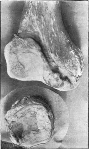
Fig. 296. - Lower end of femur and patella in chronic rheumatic arthritis. In the patella the cartilage is eroded and partly replaced by an enamel-like surface. In the femur there is an enamel-like surface replacing cartilage (especially on the left), and there is also new-formation of bone, giving the striking "lipped" appearance shown.
The further changes are the result of the wearing down of the articular ends where these are exposed to friction and at the same time the new formation of bone at the borders of the articular surfaces. These two features are shown in Fig. 296, which represents the lower end of the femur and the patella. The altered cartilage, which has a furry aspect, being worn away, the cancellated bone which would otherwise be exposed gets covered with a smooth, polished, enamel-like layer which takes the place of the cartilage. This peculiar porcelainous surface is localized at the parts which grind against each other, and there will usually be similar areas on the opposing bones of a joint.
At the edges of the articular surface the new-formation of bone sometimes, as in Fig. 296, takes the form of an expansion of the articular surface, the appearance suggesting the impression that the bone had overflowed outside the proper surface. The articular surfaces may thus have a ring or everted lip (see Fig. 296) of new bone, and the surface of this may also have the porcelainous character. This newformation of bone at the edges of the joint-surfaces is, according to Ranvier, largely from cartilage, which in the protected position at the edges undergoes proliferation and leads on to the formation of bone. There is also formation of bone in the periosteum and even in the ligaments, so that an irregular fringing of the joint with long projections may occur.

Fig. 297. - Deformity of hand from chronic rheumatic arthritis. (Robert Adams).
With all this there is considerable thickening of the ligaments by inflammatory new-formation of connective tissue, and often fibrous union between opposing parts of the joints. Indeed, if the joints are kept at rest, there may be a complete union of the parts around the joints opposite each other, leading to anchylosis. Without anchylosis there is stiffness of the joints, whose movements are greatly curtailed.
The process of grinding down of the bones along with new-formation often leads to great alterations in form of the articular ends, and great deformity in the parts concerned, as is shown in the hand in Fig. 297. This is particularly the case in the more extreme and localized forms, to which the name of arthritis deformans is more particularly given.
An extreme degree of arthritis deformans is sometimes seen in the hip joint, where it gets the special name of Morbus coxae senilis. Here the wearing down of the head of the bone is sometimes very extreme, so that ultimately the articulating surface may lie between the trochanters. As new-formation of bone occurs simultaneously at the borders of the articular surface, a kind of artificial head is produced, and the appearance is presented as if the neck were atrophied and the head displaced as in Fig. 298. In like manner an apparent widening of the acetabulum may occur. The original articular surface is worn away, but by the formation of new bone under the periosteum around, a wall is formed, giving the appearance of the borders of a widened acetabulum.
Charcot's Disease
This name is applied to conditions of the joints arising in consequence of diseases of the spinal cord, especially locomotor ataxia. The lesions consist in an atrophy of the articular ends of the bones including the cartilages. There is in consequence of the exposure of the bone, a wearing down of the bones, without the new-formation such as appears in chronic rheumatic arthritis. (See under Locomotor Ataxia).
Literature
Rheumatic Arthritis - Robert Adams, Treatise on rheumatic gout, etc., with atlas, 1857 and 1873; Lane, Trans. Path. Soc, xxxvii., 1886; Canton, ibid, iii., 1851, xii., 1861; Hutchinson, ibid., xxiii., 1872; Wilks, Guy's Hosp.
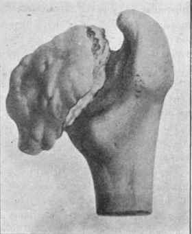
Fig. 298. - Head of femur in chronic rheumatic arthritis - the head and neck worn down and the appearance of a new head produced by new-formation. (Robert Adams).
Rep., iv., 1858; Zikglee, Virch. Arch., lxx., 1877; Weichselbaum, ibid., lv., 1872; Charcot, Senile and chronic dis., New Syd. Soc, 1881. Charcot's Disease - Charcot, Dis. of nerv. syst., New Syd. Soc, 1881.
Continue to:
My Books

