


7. Affections Of The Pleura In Phthisis
Description
This section is from the book "A Manual Of Pathology", by Joseph Coats, Lewis K. Sutherland. Also available from Amazon: A Manual Of Pathology.
7. Affections Of The Pleura In Phthisis
From the intimate connection of the pleura with the lung it may be expected that it will frequently be affected in phthisis.
The lymphatic system of the lung does not apparently communicate directly with the cavity of the pleura, so that although a tuberculosis may extend to the subpleural tissue it does not directly affect the pleural sac.
The relations of the pulmonary lymphatics to the pleura are shown by the locality of the carbonaceous pigment in the lungs. This carbonaceous pigment is carried about and deposited wherever there are communicating lymphatics, and it is often abundant in the subpleural tissue; but it never penetrates into the sac itself. On the other hand, there seems to be a communication in the opposite direction from the pleura to the interlobular connective tissue. This is shown by the fact that in tubercular pleurisy there is a certain penetration from the pleura into the lung. (See under Tubercular Pleurisy).
While tuberculosis does not extend to the pleura, because it implies the passage of solid bodies (the bacilli), there are very commonly simple inflammatory processes, which depend on the extension of the dissolved products. Such products being present in the connective tissue of the lung may readily soak into the pleura.
Chronic pleurisy is a constant occurrence, giving rise, as in other cases of chronic inflammation, to new-formation of connective tissue. The pleura over a tubercular lung is nearly always thickened, sometimes greatly so (as in Fig. 369), and the two layers are almost constantly adherent. The two layers are not only adherent, they have really coalesced, and their blood-vessels intercommunicate, so that if the vessels on either side be obstructed the pleura may be still nourished from the other. The thickening and adhesion are conservative processes, shutting off the diseased lung from the general pleural sac. It is when these conditions fail to occur that we commonly have the more serious pleural lesions, acute pleurisy and pneumothorax.
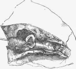
Fig. 375. - Aneurysm in a bronchiectatic cavity. A probe has been introduced into the artery, and is visible through the gaping aperture in the aneurysm. It is also indicated close to the wall of the bronchus beyond the cavity.
Acute pleurisy is a frequent concurrent in cases of phthisis, especially in the caseous form. It implies that the lesion in the lung has come to the surface at a place where adhesion of the pleura has not yet taken place. This will be most frequent in early periods and in acute cases.
The acute pleurisy is often connected with Necrosis of the pleura. The pulmonary pleura is nourished by the vessels of the lung, and as these are occluded and necrosed when involved in the caseating lesion, the pleura will be involved in the necrosis in so far as it is related to the occluded vessels. At the very outset of a caseous phthisis one of the affected areas may be immediately beneath the pleura, and we may have a necrosis before there has been time for the formation of adhesions. In this way we may explain many of the cases in which pleurisy has apparently but not really preceded the pulmonary disease. Again, in all acute cases we are liable to have necrosis of the pleura. and it is not uncommon to find quite a number of dead white areas visible on the surface, each indicating an area of necrosis, generally concealed to some extent by a layer of fibrine, the result of the inflammation.
The mere exposure of a necrosed piece of pleura seems to induce an acute pleurisy, perhaps by allowing the penetration of irritating juices such as the living structures intercept. The acute pleurisy is of the usual kind (see further on) accompanied by fibrinous exudation, but it is usually limited by existing adhesions. Through time the acute inflammation subsides and a chronic pleurisy, with adhesion, result-.
Pneumothorax also implies necrosis of the pleura, but there is, in addition, a partial separation of the dead piece and the establishment of a communication between the pleural sac and the air passages. This infers the existence of a cavity beneath the necrosed piece of pleura and not merely a caseous area. The necrosed piece begins to separate at one edge, and then the pneumothorax suddenly occurs. As death often occurs in consequence of a pneumothorax, one not infrequently has the opportunity of observing the condition in this stage. If the patient survive, then the whole piece is detached and apertures are left in the pleura such as those shown in Fig. 37G.
Along with pneumothorax there is an acute pleurisy. If it has not occurred in consequence of the necrosis, then it will ensue when, by the separation, some of the contents of the pulmonary cavity pass into the pleural sac. These two conditions, acute pleurisy and pneumothorax, are thus closely related, both of them being connected with necrosis of pleura. They also occur in relation with the advanced outposts of the pulmonary lesion, and are hence liable to develop in connection with the less affected lung or portion of lung, and by causing serious damage to the lung on which the patient chiefly depends for respiration, they often induce very serious and even fatal dyspnoea.

Fig. 376. - Lower part of lung and pleura from pneumothorax. There are three apertures caused by necrosis of pulmonary pleura.
Continue to:


My Books

