II. Coagula In The Heart
Description
This section is from the book "A Manual Of Pathology", by Joseph Coats, Lewis K. Sutherland. Also available from Amazon: A Manual Of Pathology.
II. Coagula In The Heart
Thrombi in the heart are of frequent occurrence; they vary in kind and in significance. Most of the forms of thrombi have been already referred to (see p. 95). Thrombi are frequently designated vegetations, but it is not advisable to use this word in the place of the more accurate one thrombi. We may distinguish three forms of thrombi: warty, globular, and polypoid.
Warty thrombi occur in acute endocarditis, owing to the coagulation of the fibrine on the inflamed and roughened surfaces. (See further on).
Globular thrombi are of common occurrence in dilated and hyper-trophied hearts, originating in the retired parts of the cavities, such as the auricular appendages, the apices of the ventricles, and behind the columnse cornea?. (See Figs. 204 and 205.) They are usually multiple, and the smaller of them may appear as pearly white bodies presenting a rounded projection between the trabecular They may, however, grow out from these positions, and assume considerable dimensions. It is not uncommon to findtthem .distending the auricular appendage, and sometimes filling the greater part of the auricle. The larger ones soften in the centre, forming a whitish or brownish juice, resembling pus in its naked-eye characters, but consisting merely of debris. It is quite common to find the thrombus converted into a sac, composed of a thin rind and a cavity filled with this puriform fluid. Rupture may occur during life and pieces of the thrombus may he torn off, or a thrombus may be detached bodily and carried into the pulmonary artery or aorta, so as to produce embolism. The globular thrombi are most common in the right auricle and ventricle, as these cavities are more liable to dilatation than the left, hence embolism is more frequent in the lungs than in the systemic system. The formation of thrombi in the right cavities often coincides with thrombosis in the veins, as causes which induce dilatation of these cavities are similar to those which lead to general venous hyperemia. Hence in a particular case of pulmonary embolism it may be a question whether the source is in the right heart or in the veins.
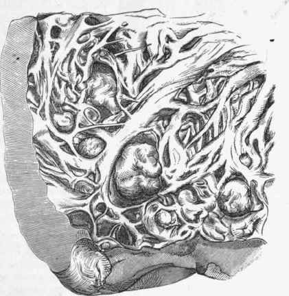
Fig. 204. - Globular thrombi near the apex of the left ventricle. Several of these are seen to project from between the mnsculi papillares.
The Polypoid thrombi are much more uncommon than the other two forms. It sometimes happens that a thrombus is formed on a valve or on the internal surface of the heart and from this point grows out by successive deposition to a considerable size. The author has met with a case in which the left ventricle was filled with massive festoons thus formed, and great hypertrophy and dilatation had occurred. In this case also the coagula had undergone a partial impregnation with lime. There is also a case recorded by Gftirdner in which a thrombus, formed of firm fibrihe and attached to the wall of the right auricle, hung free in the auricle and assumed a nearly globular form (see Fig. 205). It was so placed as to hang down into the tricuspid orifice, which it greatly obstructed, like a ball-valve.
A somewhat similar case is recorded by Allan Burns as having occurred in the Royal Infirmary, Glasgow.
In the first of the cases referred to above, the left ventricle was occupied by a large firm fibrinous mass, which consisted of twelve or thirteen polypoid coagula having a firm attachment to the anterior wall of the ventricle at about its middle. It was estimated that these coagula weighed two ounces. The coagula were obviously old and the basal parts had undergone a kind of fibrous transformation resembling tendon, while the red part looked like the fleshy part of a muscle. There was no softening, but in the substance of the polypus and partly on its surface, a somewhat massive deposition of lime salts had occurred, forming at one place a sort of stem an inch in length. (The specimen is in the museum of the Royal Infirmary, Glasgow).
The second case is one in which a stenosis of the tricuspid valve had been diagnosed by Sir William Gairdner many years before death. (This unique specimen is in the museum of the Western Infirmary, Glasgow).
Literature
Allan Burns, Dis. of heart, 1809; Steven and Coats, Glasg. Med. Jour., Feb., 1870; Gairdner, Clin. Med., 1862, and Edin. Hosp. Eeports, 1893; Wickham Legg (Loose balls of fibrine in left auricle), Path, trans., xxix., 49.
Continue to:
My Books

