Carcinoma. Malignant Tumor Of Epithelial Origin
Description
This section is from the book "A Manual Of Pathology", by Guthrie McConnell. Also available from Amazon: A Manual Of Pathology.
Carcinoma. Malignant Tumor Of Epithelial Origin
A carcinoma is a malignant tumor of epithelial origin. It is characterized by a marked proliferation of epithelium with infiltration into the surrounding tissues.
The epithelium is arranged atypically in a supporting framework made up of adult connective tissue.
The epithelial cells are not characteristic of the growth, but they differ in some respects from the normal type. The diagnosis of carcinoma cannot be made from the cell, as there is no distinct cancer cell. The general arrangement of epithelium and connective tissue must be taken into consideration.
The carcinomatous epithelium frequently consists of cells many times larger than normal. Their nuclei may be unusually large, vesicular, and show a peculiar affinity for nuclear stains, a condition called hyperchromatosis.
They may divide by an atypical mitosis and give rise to peculiar arrangements of the chromatin. These cells multiply rapidly, and, though at first round, they may become almost any shape on account of the mutual pressure exerted.
In some cases giant cells occur. Tumors of this variety differ greatly in size, shape, color, and density.
Carcinomas are composed of two types of tissue, epithelial and connective, cells and stroma. According to the one that predominates, carcinoma are called medullary when the cells are more numerous; scirrhus when the tumor is rich in connective tissue.

Fig. 60. - Carcinoma of Mammary Gland (Mallory).
Medullary type of growth. Slight tendency to the formation of gland lumina.
The first is soft; the second, hard.
Well-developed blood-vessels and lymphatics are found in the stroma, which is most likely derived chiefly from preexisting connective tissue, but a certain amount is probably newly formed. Elastic fibers are present in the infiltrating portion of the growth, but they are fragments of fibers preexisting in the invaded tissue. The cellular elements originate from the epithelium normal to the part involved, and frequently retain the characteristics of the primary cell.
The more closely connected it is with the original cell, the more does the carcinoma cell resemble it. The further away it is, the greater is the variation. There is then a tendency to revert to the round, undifferentiated, embryonal type. Between the cells no fibrillary substance is found.
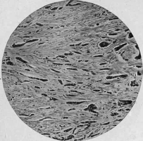
Fig. 61. - Scirrhous Carcinoma of Breast (Mallory). Alveoli of epithelial cells small; stroma abundant.
In carcinoma the cells frequently undergo degeneration, and usually of a form peculiar to the parent tissue. If it arose from squamous epithelium, keratin is found; colloid or mucoid material if derived from mucous membranes. The tumor may break down and undergo a fatty change, most common in the mammary gland.
A carcinoma may become infected and show marked inflammatory changes which may be so great as to somewhat disguise the true character of the growth. There will be an infiltration of the tissues with leukocytes.
Microscopically a carcinoma consists of columns of cells running in all directions, separated from one another by fibrous tissues. These columns give the appearance of alveoli filled with epithelium. The columns are branched into numerous subdivisions, giving a complicated root-like structure.

Fig. 62. - Carcinoma of Mammary Gland (Mallory and Wolbach). Extension of tumor through a lymphatic in fat tissue.
As the tumor grows these cells infiltrate and ramify in all directions, occupying usually the lymphatic spaces. Along the advancing border there is a more or less well-marked zone of round-cell infiltration.
As there is no intracellular substance, the cells easily break away from the main mass and are carried to the neighboring lymph-nodes. This may take place very early and give rise to extensive metastasis. These secondary growths are usually similar in character to the primary.
Extension to distant tissues may be due to permeation.

Fig. 63. - Squamous Epithelioma (McFarland).
a, Epithelial masses; b, epithelial pearls; c, connective tissue; d, capillary blood-vessels.
According to this theory there is a continuous growth of cancer cells along the lymphatics of the deeper fascia, with widespread involvement.
Continue to:
My Books

