Odontoma
Description
This section is from the book "A Manual Of Pathology", by Guthrie McConnell. Also available from Amazon: A Manual Of Pathology.
Odontoma
An odontoma, according to Bland-Sutton, from whose work the following is taken, is a tumor composed of dental tissues in varying proportions and different degrees of development, arising from the teeth germs, or teeth still in the process of growth.
The species of this genus are determined according to the part of the tooth germ concerned in their formation.
1. Epithelial odontoma = from the enamel organ.
2. Follicular odontoma,
3. Fibrous odontoma,
4. Cementoma,
5. Compound follicular odontoma, = from the tooth follicle.
6. Radicular odontoma = from the papilla.
7. Composite odontoma = from the whole germ.
1. Epithelial Odontoma
Epithelial Odontoma occur, as a rule, in the mandible, but they have been observed in the maxilla. They have a fairly firm capsule, and in section display a collection of cysts of various shapes and sizes, but the openings rarely exceed 2 cm. in diameter. The cysts are separated by thin fibrous septa, sometimes ossified. The cavities contain a brown mucoid fluid which gives a.reddish tint to the growing portions of the tumor. Histologically, an epithelial odontoma consists of branching and anastomosing columns of epithelium, portions of which form alveoli. The cells occupying the alveoli vary, the outer layer may be columnar, while the central cells degenerate and give rise to tissue resembling the stellate reticulum of an enamel organ. It may be that many of these tumors do not arise from the epithelial enamel organ, but from endothelium within the gums.
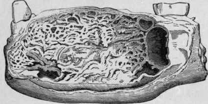
Fig. 54. - Epithelial Odontoma. Natural Size (Bland-Sutton).
2. Follicular Odontoma
Follicular Odontoma comprise those swellings often called "dentigerous cysts," an inaccurate term. They arise commonly in connection with teeth of the permanent set, and especially with the molars. Sometimes they attain large dimensions and produce great deformities, particularly when they arise in the upper jaws and happen to be bilateral. Occasionally they occur in connection with supernumerary teeth. The tumor consists of a wall of varying thickness, which represents an expanded tooth follicle; in some cases it is thin and crepitant, in others it may be as much as I cm. thick. The cavity of the cyst usually contains viscid fluid and the crown or the roof of an imperfectly developed tooth. Occasionally the tooth is loose in the follicle, sometimes inverted, and often its root is truncated. Exceptionally the tooth is absent, or is represented by an ill-shaped denticle. The walls of the cyst always contain lime or osseous matter, the amount varying considerably. These tumors are not unknown in other animals, having been found in sheep, pigs, and porcupines. The amount of fluid in a follicular odontoma varies, and the size of the tumor depends in the main upon this. Sometimes the fluid may measure as much as 2 ounces, and this may lead to the wide separation of the inner and outer plates of the body of the mandible, and the odontoma may occupy the entire length of the bone.
3. Fibrous Odontoma
In a developing tooth, a portion of the connective tissue in which it is embedded is found to be denser and more vascular than the rest; it also presents a fibrillar arrangement. This condensed tissue is known as the tooth sac, and, when fully developed, presents an outer firm wall and an inner looser layer of tissue. At the root of the tooth the follicle wall blends with the dentin papilla, and is indistinguishable from it. Before the tooth cuts the gum it is completely enclosed within this capsule. Under certain conditions this capsule becomes greatly increased in thickness and so thoroughly encysts the tooth that it is never erupted. Such thickened capsules are mistaken for fibrous tumors, especially if the tooth be small and ill developed. Under the microscope they present a laminated appearance, with strata of calcareous matter. To these the term "fibrous odontomata" may be applied. As a rule they are multiple, four being by no means an unusual number. There is good reason to believe that rickets is responsible for some of these thickened capsules.
4. Cementoma
When the capsule of a tooth becomes enlarged, and these thick capsules ossify, the tooth will become embedded in a mass of cementum. To this form of odontoma the name "cementoma" may be applied. Tumors of this character occur most frequently in horses. The chief structural peculiarity is the presence, in enormous numbers, of large, richly branched openings.
5. Compound Follicular Odontoma
If the thickened capsule ossifies sporadically instead of uniformly, a curious condition is brought about, for the tumor will then contain a number of small fragments of cementum, or dentin, or even ill-shaped teeth (denticles) composed of three dental elements - cementum, dentin, and enamel. The number of teeth or denticles varies greatly, and may reach a total of four hundred.
6. Radicular Odontoma
Radicular Odontoma is the term applied to those tumors which arise after the crown of the tooth has been completed, and while the roots are in the process of formation. As the crown of the tooth, when once formed, is unalterable, it naturally follows that should the root develop an odontoma, enamel cannot enter its composition; the tumor would consist of dentin and cementum in varying proportions, these two tissues being the result of the activity of the papilla. The outer layer of the odontoma is composed of cementum; within this is a layer of dentin, and inside this is a nucleus of calcified pulp. It is probable that some radicular odontomata in man are due to inflammatory changes.
7. Composite Odontoma
Composite Odontoma is a convenient term to apply to those hard tooth tumors which bear little or no resemblance in shape to teeth, but occur in the jaws, and consist of a disordered conglomeration of enamel, dentin, and cementum. Such odontoma may be considered as arising from an abnormal growth of all the elements of a tooth germ, enamel organ, papilla, and follicle. Not only is this growth composite in that the tumor originates from all the elements of a tooth germ, but it is composite in another sense: many of these tumors are composed of two or more tooth germs indiscriminately fused. But they differ from the cementomata containing two or more teeth in the fact that the various parts of the teeth composing the mass are indistinguishably mixed, whereas the individual teeth implicated in a cementoma can be clearly defined. It was long believed that composite odontomata occurred only in the mandible, but it is clear not only that they arise as frequently in the maxillae, but that they attain a far larger size in the upper than in the lower jaw.
Continue to:
My Books

