Psammoma
Description
This section is from the book "A Manual Of Pathology", by Guthrie McConnell. Also available from Amazon: A Manual Of Pathology.
Psammoma
Psammoma is a tumor allied to the sarcoma. It is made up of masses of spindle cells, which contain areas of hyaline degeneration and calcification. Are usually found in the meninges of the brain and spinal cord. Endothelioma is a tumor arising from endothelial cells. These growths are at times very difficult to differentiate from carcinoma on account of the apparent cell nest arrangement, but careful examination will commonly show some sarcomatous areas.
The cells extend along the lymphatic spaces and are closely related to connective tissue. Are found in the serous membranes, testicle, ovary, liver, and parotid. Are malignant.
Under this same heading are included those tumors developing from the cells in the lymph-spaces, the lymphangio-endo-thelioma; and those from the endothelium of the blood-vessels, the hemangio-endothelioma. Occasionally some of these tumors arise from the perivascular endothelium, and to these has been given the name perithelioma. The cells in these latter are arranged in strands radiating from the vessel around which the mass originated.
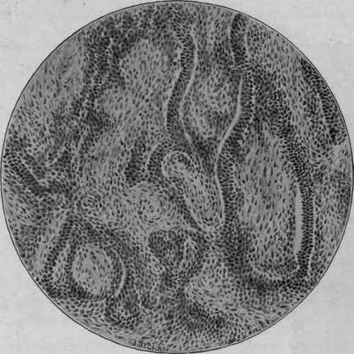
Fig. 46. - Endothelioma of the Pleura. Zeiss, Oc. 2; ob. c.
(McFarland).
The illustration shows the cellular growth in the form of cylindric masses which fill crevices of the tissue, probably originally channels.
According to the combination of tissues present sarcomata may be further classified as follows: all such varieties, however, not being mentioned:
Osteosarcoma = bone present.
Chondrosarcoma = cartilage present.
Myosarcoma = muscle present.
Neurosarcoma = nerves present.
Tumors Of Adult Connective Tissue
Fibroma is a tumor of fibrous connective tissue. Fibromata are usually pale in color, round, lobulated, circumscribed, and encapsulated. They may be of varying degrees of firmness.
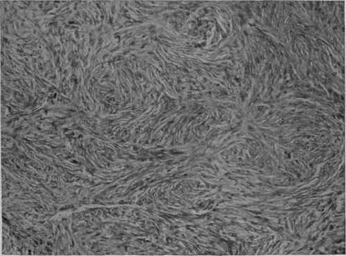
Fig. 47. - Fibroma. Cells and Fibrils in Small Bundles which Run in Every Direction (Mallory).
The cells resemble those of normal connective tissue and are arranged in bundles that cross each other in all directions.
In the soft variety the cells are separated by serous or mucous deposits.
In the hard variety the cells are closely packed together.
Continue to:
My Books

