Syphilis
Description
This section is from the book "A Manual Of Pathology", by Guthrie McConnell. Also available from Amazon: A Manual Of Pathology.
Syphilis
Is a specific, infectious, and very contagious disease of man. By experimental inoculation it has been transmitted to the higher apes and rabbits.
Is due to the Treponema pallidum (Spirochceta pallida), an organism that has been so constantly found in syphilitic lesions that it seems most probable that it is the causative factor. This organism is long, actively motile, spiral or corkscrew in shape, and with pointed ends. It is from 4 to 20. long, 0.25, thick, and contains six to fourteen turns which are short, clear cut and regular. It is difficult to stain and to grow, but it has been obtained in pure culture.
The disease may be divided into the -
1. Period of primary incubation, about three weeks.
2. Period of primary symptoms, chancre and adenitis.
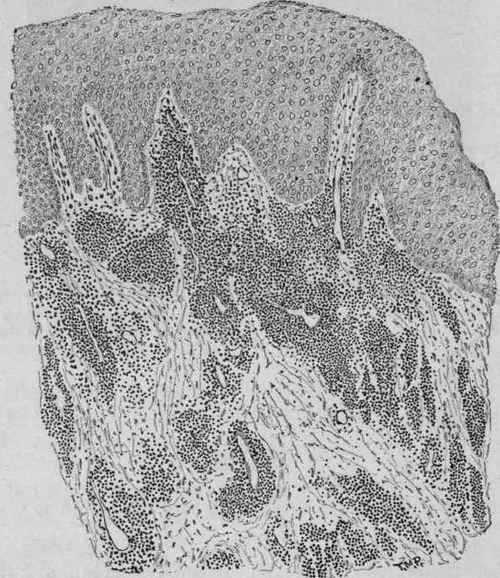
Fig. 36. - Section from a Primary Syphilitic Nodule of the Mucous Membrane of the Mouth, Showing Collections of Cells About the Blood-vessels in the Submucous Tissue (Delafield and Prudden).
3. Period of secondary incubation, about six weeks.
4. Period of secondary symptoms, from one to three years.
5. Intermediate period of two to four years, during which the patient may recover.
6. Period of tertiary symptoms, unlimited.
The primary lesion is the chancre, which starts as a single papule, seldom multiple, at the seat of inoculation, which may be either genital or extragenital, and is invariably present except in congenital syphilis. This soon becomes eroded as a result of superficial necrosis, but increases in size due to infiltration of the deeper tissues. Is circular or oval, 1 by 1.5 cm., base hard. The edges are sharply defined, the induration not extending much beyond the lesion, and terminating abruptly. The chancre is slightly elevated and movable, not being adherent to the underlying tissues. Secretion is thin and scanty, suppuration being unusual; the surface may be dry or covered by a slight false membrane.
Microscopically there is a tremendous infiltration of cells, particularly in the neighborhood of the vessels, and marked changes in the walls of the blood-vessels. The infiltration begins with an escape of small round lymphoid cells, and this is accompanied by a proliferation of the cells of the cutaneous connective tissue and the cells of the walls of the blood-vessels. The elastic tissue disappears, the new formation of cells extending along the small arteries and veins. The tissue becomes crowded with various kinds of cells - polymorphonuclear leukocytes, lymphocytes, plasma cells, endothelial and connective-tissue cells, and fibroblasts. The media of the vessels thickens, the endothelium of the intima proliferates, and the lumen may be partially or completely obstructed. As a result of the interference with the circulation degeneration begins. The vessels not infrequently show hyaline changes.
Continue to:
My Books

