Microscopical Examination Of The Gastric Contents. Part 2
Description
This section is from the book "Diseases Of The Stomach", by Max Einhorn. Also available from Amazon: Diseases of the Stomach.
Microscopical Examination Of The Gastric Contents. Part 2
(C) Mould Pellicles
The part which micro-organisms (bacteria and mould fungi) play in the occurrence of pathological processes in the stomach has been variously interpreted by authors. Most clinicians ascribe no special significance to them. Others, however, assign them a prominent place; thus, for exampie, Talma1 maintains that the fermentation of carbohydrates induced by micro-organisms is the cause of hyperchlorhydria; others, again, place stress not so much upon the variety of these microbes as upon their ultimate number. Among these authors Naunyn' may be especially cited.

Fig. 22. - A Specimen of Mucus from the (Esophagus (from a Patient with Carcinoma Cardiae, J. C. W.), showing mucus, bacteria, fat and epithelial cells, some of the latter grouped together.
1 Talnm: "Von der Gillmmg der Kohlekydrate im Magen." Zeit-schr. fur klin. Mudicin. 1898. Bd. 35, p. 542.
2 B. Naunyn: "Uebcr das Vehaltniss der Magengahrungen." Deutsches Arch. f. klin. Med., vol. xxxi.
The mould fungus, as such, has been but little mentioned in the domain of gastric affections.
In all the literature the scant references to mould fungi in the stomach relate to the findings of microscopical examinations. Mould itself, recognizable by macroscopic examination, has, according to my knowledge, not as yet been observed clinically in the stom-ach. At any rate, no mention of this occurs in the literature. Il have had occasion to observe several cases of mould formation in the stomach. In the cases under my observation there were found in the wash water of the empty stomach small, sometimes blackish-gray, and sometimes brownish-green flakes (2 to 5 mm. 'in diameter; see Fig. 23) in varying number (four to fifty and more). The microscopical examination showed that these flocculi consisted entirely of spores and mycelia and scarcely anything else. Similar flocculi were found in the same patients in the gastric contents after a test meal, and the microscope showed the same picture as in the flocculi from the empty stomach.

Fig. 23. - Small Pellicles of Mould found in the Stomach. (Natural size).
1 Max Einkorn: "The Occurrence of Mould in the Stomach and its Probable Significance." Medical Record, June 16th, 1900.
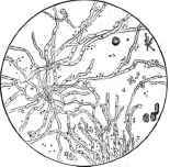
Fig. 24.- A Greenish Pelliele found in the Wash-water of the Stomach (of Wm.R-----). in yhe fasting condition. Mycelia, free spores, and a few crystals are visible. X 340.
Sometimes these blackish-gray masses are embedded in mucus. We then note besides these fungus colonies mucous corpuscles and numerous epithelial cells. This indicates an intimate connection between the fungus colonies and the surface of the mucous membrane. The former must adhere quite closely to the latter and perhaps even proliferate into the epithelial layer. This firm adhesion must bo assumed for the following reasons: If the fungi were only an accidental admixture of the ingesta, that is, introduced with the latter and then carried farther onward, without there being any fungus proliferation, then they would be encountered only in the gastric contents.
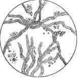
Fig. 25. - Smile us Fig. 24, hlghly magnified. X 420.
That the grayish-green or grayish-black flakes, which were found, represented mould pellicles, was established beyond doubt by the microscopical examination. An extremely large number of spores and mycelia were always observed. In all my cases the microscopical picture was the same, and it can therefore be assumed that the mould fungi present belong to one and the same species. Dr. E. K. Dunham has identified them as penicillium glaucum.
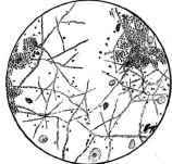
Fig. 28. - A Blackish Pellicle found in the Gastric Contenta (of T. M-----) alter a Test Breakfast. Numerous spores, micella, a tew crystals, standi granules, and epithiellal velbs are visible.
What significance have these mould fungi in gastric pathology? Although isolated fungi may exist in the stomach for a short time without any detriment, they do not find in the normal organ favorable soil for further development. They are intimately mixed with the chyme and are carried onward, living or dead, through the pylorus. Entire colonies of fungi which are macroscopically perceptible are probably never to be found in the normal stomach. Any considerable growth of mould would be possible only if a colony of the fungi had infested a fold of the surface of the gastric mucous membrane and had become so firmly adherent that they were not carried along with the onward passage of the chyme. Under these circumstances a fungus colony may grow undisturbed, and considerable areas of the gastric mucosa may become covered with mould. In my cases such a condition must have prevailed. In lavage of the stomach the inflowing current of water exerts considerable force and tears many mould islets from their bases, so that they then appear in the wash water.
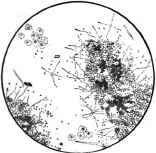
Fig. 27. - A Blackish Pellicle found in the Wash-water of the Stomach (L. C----). In the luting condition. Numerous spure-colonies, mycella, a few crystalk, epithelial cells, and several algae are vlssltile. X 120).
It is scarcely conceivable that such a mould coating of certain zones of the gastric mucosa can be unattended with disturbances of the functions of the organ. Conditions of irritation as well as inflammatory processes might be expected a priori from the mechanical action of the mould.
After these theoretical conclusions it would be profitable to analyze more closely the cases observed, and to elucidate whether the mould formation was in a causative relationship to the symptoms of the disease. The decision of this question is, however, very difficult, because post hoc is not always ergo propter hoc. I have met with the mould formation particularly in two groups of gastric affections: first, in cases of intense hyperchlorhydria (occasionally attended with hypersecretion and vomiting); and, second, in gas-tralgia with normal or reduced gastric secretion. It cannot be denied that in many of these cases the mould flakes became smaller in number or disappeared after gastric lavage followed by spraying with a one to two per mille solution of nitrate of silver. In connection with this a subjective improvement could be observed in the condition of the patient. Yet it cannot be said with certainty that the mould produced the existing pathological process in the stomach; for we find cases analogous in every respect without the presence of mould fungi.
Notwithstanding this, it appears plausible that these mould fungi are connected to a certain extent with the above-mentioned abnormal conditions; and even if they are not the cause of these, they undoubtedly increase their severity.
The occurrence of mould in the stomach in large masses must, therefore, be considered of importance from a therapeutic standpoint; hence it must be our endeavor to free the stomach from them as soon as possible. This is best done by irrigation of the stomach in the fasting state of the patient. This acts in a purely mechanical manner, since the mould flakes are removed with the water. The use of the gastric douche might also have a favorable influence in this direction. Following this the application of an antiseptic solution of silver nitrate with a spray appeal's likewise of some utility. Aside from the therapeutic measures just described, the treatment of these cases must be directed in accordance with the special disease present.
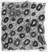
Fig. 38. - Group N (Nornal). A small piece of gasirie muensa (patient Mrs. H.) presenting a cross-sectlon of the glands in normal appearance. X 80.
Continue to:
My Books

