


Microscopical Examination Of The Gastric Contents. Part 3
Description
This section is from the book "Diseases Of The Stomach", by Max Einhorn. Also available from Amazon: Diseases of the Stomach.
Microscopical Examination Of The Gastric Contents. Part 3
(D) Small Pieces Of Gastric Mucosa
In washing out the stomach (especially in the fasting condition) occasionally a small piece of gastric mucosa may be found in the wash-water. Such a small piece of gastric mucosa may also be found occasionally in the gastric contents when examining the patient after a test breakfast or test dinner. Boas1 was the first to make use of such specimens for microscopical examination. He was of the opinion that such an examination permits one to judge of the morbid anatomical condition of the given case. A short time afterward I observed that in some cases the occurrence of small pieces of gastric mucosa in the wash-waier is a constant phenomenon.
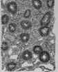
The number of these pieces varies from one to four (see Erosions of the Stomach). During the last ten years I had the opportunity to examine a great number of such small particles of gastric mucosa, a large part of which belonged to cases of erosions of the stomach, the remainder to many other affections. Such a piece of gastric mucosa looks quite red. The thickness may vary from 1/2 to 1 mm., while the size may vary from that of a large pin's head to that of a small bean. Sometimes they are found embedded in mucus. While the presence of glands in these small pieces may be found by examining them in the fresh condition under the microscope, a thorough examination can he made only after a sufficient preparation of these particles (hardening in alcohol, embedding in celloidin and staining with eosin, hematoxylin, picro-carmine, methylene blue, and thionin).
1 J.Boas:L.c., p. 225.
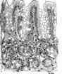
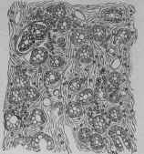
In examining the microscopical picture of the different specimens the following groups can be easily distinguished: 1. N = Normal: glands and interglandular tissue exist in normal proportions. 2. C = Connective tissue: while there is a normal proportion between glands and interglandular tissue. there is a marked proliferation of connective tissue around the glands.
3. P = Proliferation: there is a marked proliferation of glands; they are nearer each other and sometimes have an elongated and curved shape.
4. B = Beginning Atrophy: the glands exist In smaller numbers, and are sometimes also smaller in size; the interglandular spaces being quite large and filled partly with small-cell infiltration partly with connective-tissue formation. 5. A = Atrophy: complete atrophy; no glands visible only indications of their previous existence; round-cell infiltration.
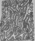
6. V: Vacuolization: within the glands exist vacuoles of different shape, being the result of a mucoid degeneration of some glandular cells.
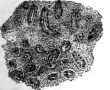
Fig.32. - Group B (Beginning Atrophy). A piece of gastric mucosa (from patient B. E. situ carcinoma cardiae), showing construction of glands by connective-tissue proliferation.
Sometimes one specimen shows characteristics be-longing to two of the groups mentioned. For the beautiful execution of the drawings I am indebted to Dr. C. A. Elslierg. who made them from my specimens (see Figs. 23 to 34). Although I think that the microscopical examination of these pieces of gastric mucosa is of great interest and may occasionally help to supplement the diagnosis I do not believe that it permits us to judge positively about the original affection of the stomach, for in some cases I have noticed in the microscopical picture very few small glands, the whole field having the appearance of atrophy, and still the gastric secretion was perfectly normal. On the other hand, I' had a patient with distinct symptoms of chronic of tissue may be found. These, if examined under the microscope, may occasionally reveal the nature of a tumor, whether cancerous or not. The examination is of importance if a characteristic picture of a malignant type is discovered. Most frequently such pieces may be obtained in cases of cancer of the cardia.
I append a drawing obtained from a specimen of such a small piece of cancerous tissue from a patient with cancer of the cardia (Fig. 35).

Fig. 33. - Group A (Atrophy). A piece.of gastrle from patient R. H. D). No glands visible, only some empty spaces where glands had previously existed..
1 For further details see Max Einhorn: "The State of the Gastric Mucosa in Secretory Disord em of the Stomach," Medical Rec-oid, June 27th, 1896 gastric catarrh and diminished gastric secretion in whom the pieces of gastric- mucosa found in the wash-water presented a perfectly normal appearance (Fig. 21).
(E) Particles Of Tumors
In the gastric contents obtained after test meals, in the vomited matter, in the wash-water after lavage of the stomach, or within the tube after an exploratory examination, small particles and was thicker and firmer than pieces of gastric mucosa. Cross-section presents all appearances of alveolar carelnoms x 100.
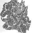
Fig. 35 - A piece of Tumor (from B. E.)Obtained after Examination with Stomach Tube. Id fresh condition it appeared white.
Continue to:


My Books

