Clinical Features Of Cancer Of The Kidney
Description
This section is from the book "Early Detection And Diagnosis Of Cancer", by Walter E. O'Donnell. Also available from Amazon: Early Detection And Diagnosis Of Cancer.
Clinical Features Of Cancer Of The Kidney
Localized Lesion Symptoms
1. Blood in the urine. Gross hematuria is present at some time in about 75% of patients with kidney cancer and is the first manifestation in around 40%.
(a) Reddish or smoky discoloration is present throughout the urinary stream (total hematuria).
(b) Usually painless.
(c) May be only a single episode, subside after a few days, or persist.
(d) Pain resembling renal colic may be associated with passage of blood clots.
(e) In the case of cancer of the renal pelvis, bleeding is especially common and may be particularly profuse and persistent.
2. Pain. Some kind of pain is present with most kidney tumors, but the type and intensity are very variable.
(a) May be lumbar and/or flank and upper quadrant pain or discomfort associated with the expanding tumor itself.
(b) May be due to obstructive phenomena (e.g., hydronephrosis).
(c) May be typical pain of renal colic due to passage of clots or tumor fragments down the ureter.
3. Other symptoms. These may include the following:
(a) In the case of cancer of the renal pelvis, symptoms of chronic infection and/or stones in the urinary tract may be prominent. There may, in fact, be a long preceding history of these complaints.
(b) Anorexia.
(c) Weight loss.
(d) Nonspecific gastrointestinal symptoms.
(e) Symptoms of anemia.
(f) Fever, sweats.
The foregoing, individually or in various combinations, may be the first or only manifestation of even early renal cancer.
Signs
1. There may be no physical findings.
2. A palpable mass may be present.
(a) Presents as a firm, movable and ballotable, nontender mass in the upper quadrant of the abdomen, descending below the costal margin laterally on deep inspiration.
(b) Relatively common in cancer of the renal parenchyma.
(c) Less common to have palpable mass in early carcinoma of the kidney pelvis; occasionally, however, obstructive phenomena (e.g., hydronephrosis ) may produce this picture.
(d) Remember that the kidneys (especially the right) of thin, asthenic individuals are normally palpable.
(e) Differentiation of renal from other mass (e.g., splenomegaly) is important.
(f) Virtually impossible to differentiate clinically a mass produced by a benign renal lesion (e.g., cystic disease) from that of cancer (see Fig. 77, A and B).
3. Other signs. Much less common and prominent with a localized lesion than in the presence of a palpable mass.
(a) Evidence of weight loss.
(b) Evidence of anemia.
(c) Venous pattern over anterior abdominal wall.
(d) Varicocele.
(e) Hypertension. Occurs in about half of children with Wilms' tumors. Otherwise there is no apparent relation between kidney tumors and blood pressure.
Advanced Lesion
Almost all cases of kidney cancer exhibiting the so-called classic triad of hematuria, pain, and flank mass will prove to be advanced.
In the absence of demonstrable distant metastases, no final clinical judgment is possible on whether a given tumor is localized or advanced, operable or inoperable. This is a decision which must usually be reserved for surgical exploration.
Distant metastases may appear in the form of the following:
1. Nodular deposits in the lungs as seen on x-ray films of the chest
2. Osteolytic areas, especially in the long bones and spine and pelvis, that may present as a pathologic fracture
3. Liver enlargement and nodularity
4. Lymphadenopathy in the periaortic, mediastinal, cervical, or supraclavicular areas
Occasionally, cough, back pains, etc. may be the first or predominant symptoms or signs of the disease.

Fig. 77. A, Intravenous pyelogram that shows the left kidney to be displaced slightly downward and associated with a 15 cm. ovoid homogenous masslike density above the left kidney extending to the left leaf of the diaphragm.
B, On the retrograde pyelogram of the same patient. A, the left upper calyces are outlined and appear hydronephrotic, but the associated mass still may be clearly seen. This proved to be a rather large benign cyst of the upper pole of the left kidney.
C, Barium enema study showing an extrinsic pressure effect on the superior border of the proximal portion of the transverse colon. Note the soft tissue mass clearly defined in the right upper quadrant of the abdomen.
D, Right retrograde pyelograni of the same patient, C, shows the filling of the large kidney with hydronephrosis due to a renal cell cancer.
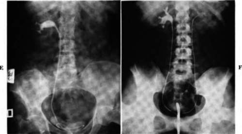
Fig. 77-cont'd. E, Right retrograde pyelograni showing a distortion of the calyces and an incomplete filling of all the calyces. The axis of the kidney is rotated. This was due to a renal cell carcinoma.
F, Right retrograde pyelograni showing a distortion of the lower calyceal group with irregularity and a loss of the normal calyceal outline. The infundibulum is widened. This is due to a cancer of the lower pole of the right kidney.
Initial Steps In The Diagnosis And Management Of The Kidney Cancer Suspect
The initial studies most commonly used and readily available to the physician are as follows:
1. Urinalysis
2. Flat film of the abdomen
3. Intravenous pyelogram
4. Cytologic examination of urinary sediment
5. Hemoglobin and/or hematocrit determination and white blood count
7. Blood urea nitrogen
At the disposal of the urologist for intensive work-up are the following:
1. Cystoscopy
2. Retrograde pyelography
Occasionally used in special circumstances by experts in the technique arc the following:
1. Aortography
2. Nephrotomography
3. Retroperitoneal air insufflation
4. Percutaneous renal biopsy
Usually resort must be made to surgical exploration and pathologic study of the kidney in question if suspicion of a tumor persists.
Urinalysis
1. Microscopic examination of the centrifuged urinary sediment for the presence of red blood cells should be a routine part of the laboratory screening of all patients.
2. If microscopic hematuria (i.e., more than 1 to 2 red blood cells per high-power field) is noted:
(a) Obtain several repeat specimens at intervals of several days.
(b) Consider possibility of:
(1) Contamination (e.g., menstruation)
(2) Hematuria due to known benign urologic disease (e.g., stones, etc.)
(3) Hematuria due to nonurologic disease or therapy (e.g., blood dyscrasias, anticoagulants, etc.)
But do not be too hasty in attributing hematuria to these less ominous problems.
(c) Remember that hematuria due to kidney cancer is more often than not microscopic and intermittent. Small assurance can be placed, therefore, on cessation of gross bleeding or a single follow-up negative urinalysis. The diagnosis of urinary tract cancer can be delayed for months unless this fact is recognized.
(d) If microscopic hematuria is not reconfirmed by several repeat urinalyses and the patient is totally asymptomatic, it may be safe to attribute the original finding to laboratory error or to benign or nonurologic disease or to label it idiopathic. Such a course of action must be taken only after serious consideration of the possibilities, however. Some physicians will elect to do an intravenous pyelogram and settle the matter.
(e) If microscopic hematuria is reconfirmed in the absence of other causes, intravenous pyelogram represents minimum further work-up. In all likelihood other studies among those listed will have to be done as well, particularly when the patient is over 40 years of age.
(f) Remember: negative urinalyses, even persistently negative ones, do not rule out the possibility of a renal tumor!
Flat Film Of The Abdomen
This may show a kidney to be abnormal as a result of any of the following:
1. Enlargement
2. Irregularity
3. Displacement
4. Calcification
Under no circumstances does a negative flat film of the abdomen rule out kidney tumor.
Intravenous Pyelogram
Visualization of the kidneys and urinary tract following the intravenous injection of contrast medium is a basic part of any urologic work-up (see Fig. 77).
In the case of renal carcinoma, the appearance of the intravenous pyelogram depends particularly on whether the lesion arises in the parenchyma (renal cell carcinoma or hypernephroma and Wilms' tumor) or in the renal pelvis (papillary or squamous types).
In general, the pyelogram may show any of the following:
1. Normal function. The renal outline, as well as the renal calyces and pelvis, may occasionally be normal in appearance despite the presence of a tumor.
2. No function. The renal calyces and pelvis may fail to be visualized.
3. Alterations in the appearance of the renal calyces and pelvis and, indirectly, the kidney parenchyma, usually in the form of:
(a) Compression
(b) Elongation
(c) Blunting
(d) Filling defect
The latter are the usual pyelography findings in the case of kidney cancer.
If suspicion of kidney tumor persists after a negative or normal intravenous pyelogram (e.g., unexplained flank pain, gross or microscopic hematuria, abdominal mass, etc.), further diagnostic studies should be done. A negative intravenous pyelogram does not rule out kidney cancer.
Continue to:
My Books

