Physical Examination
Description
This section is from the book "Early Detection And Diagnosis Of Cancer", by Walter E. O'Donnell. Also available from Amazon: Early Detection And Diagnosis Of Cancer.
Physical Examination
The complete physical examination of the apparently healthy adult can be carried out within the confines of a single ordinary examining room. Furthermore, although assistance is always helpful, especially during the pelvic and sigmoido-scopic examinations, it is possible to accomplish the entire routine without a nurse in attendance. With female patients it is usually desirable to have a nurse near by, but she can be "on call" for only portions of the examination.

Fig. 3. Equipment needed for basic cancer detection examination.

Fig. 4. Equipment needed for further work-up.
The patient should be completely undressed. The temperature, pulse rate, respiratory rate, and blood pressure, height, and weight can be determined at this time.
Some physicians prefer that the patient undergo preparation for sigmoidoscopy at this juncture. If the enema has been given at home or in the office before the doctor sees the patient, the entire physical examination can be completed without interruption. Other physicians, however, do everything but the sigmoidoscopy and then record their findings during the ten minutes or so required for bowel preparation. This is entirely a matter of personal preference and office necessity.
By and large, the physical examination is simply a very thorough check of all organ systems. It does emphasize certain areas and procedures in the search for cancer or its precursors. It is the latter aspects of the examination which will be dealt with here.
General
The general appearance of the patient should be observed especially for evidence of pallor, plethora, weight loss, jaundice, or other stigmas of ill health.
Skin
The skin in general can be surveyed at the beginning and then each area reviewed in detail as that site is covered during the course of the physical examination. The skin of the head and neck region, lips, and dorsum of the hands is most important from the standpoint of carcinoma, since about 90% of such lesions occur in these areas. Pigmented nevi are most significant on the palms and soles, in the genital region, and in areas of irritation, since these are the sites of predilection for the development of melanoma.
Lymphatics
The node-bearing areas are most conveniently examined one by one as the physician progresses from one part of the body to another. Special attention is given to the possibility of adenopathy in the cervical, submaxillary, submental, supraclavicular, axillary, and inguinal regions.
Chest
Examination of the chest consists of the conventional maneuvers of inspection, palpation, percussion, and auscultation.
Special attention is given to the presence of abnormalities of contour and expansion as well as other evidence of emphysema, atelectasis, pneumonitis, effusion, wheeze, etc.; clubbing of the fingers is looked for.
Abdomen
The usual techniques of inspection and palpation of the abdomen are carried out.
Of particular interest from the cancer standpoint, of course, is enlargement of the liver, kidneys, and spleen as well as any evidence of abdominal masses, tenderness, and distention, etc.
Male Genitalia
Careful examination, with palpation of the inguinal regions and scrotal contents, is routine.
The penis should be palpated and all skin and mucosal surfaces examined in good light. In the uncircumcised male the foreskin should always be retracted as completely as possible for inspection of the glans and coronal sulcus.
Prostate, Lower Rectum, And Anus
The prostate can be examined with the patient in the lithotomy, knee-chest, or lateral Sims' position. It also may be palpated satisfactorily while the patient is bending forward 90° from a standing position with his arms resting on the examining table.
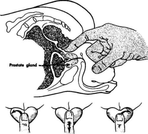
Fig. 20. Technique for examination of the prostate gland.
The posterior surface of the prostate, as well as adjacent seminal vesicles, should be reached by the examining finger (Fig. 20) and the lobes checked for irregularity of size, shape, or consistency.
The full circumference of the accessible reaches of the rectum plus the anal canal should be examined. Asking the patient to bear down during digital examination of the rectum may allow greater coverage. A stool specimen for the guaiac test may be obtainable on the examining finger at this time (p. 73).
The perirectal and presacral structures should be carefully checked during digital examination, using both thumb and index finger to feel the intervening tissue.
Continue to:
My Books

