Chapter II. Compound Microscopes
Description
This section is from the "Histology of Medicinal Plants" book, by William Mansfield. Also see Amazon: Histology of Medicinal Plants.
Chapter II. Compound Microscopes
The compound microscope has undergone wonderful changes since 1667, the days of Robert Hooke. When we consider the crude construction and the limitations of Robert Hooke's microscope, we marvel at the structural perfection and the unlimited possibilities of the modern instrument. The advancement made in most sciences has followed the gradual perfection of this instrument.
The illustration of Robert Hooke's microscope (Fig. 7) will convey to the mind more eloquently than words the crudeness of the early microscopes, especially when it is compared with the present-day microscopes.

Fig. 7. Compound Microscope of Robert Hooke.
Structure Of The Compound Microscope
The parts of the compound microscope (Fig. 8) may be grouped into - first, the mechanical, and, secondly, into the optical parts.

Fig. 8. Compound Microscope.
The Mechanical Parts
1. The foot is the basal part, the part which supports all the other mechanical and optical parts. The foot should be heavy enough to balance the other parts when they are inclined. Most modern instruments have a three-parted or tripod-shaped base.
2. The pillar is the vertical part of the microscope attached to the base. The pillar is joined to the limb by a hinged joint. The hinges make it possible to incline the microscope at any angle, thus lowering its height. In this way, short, medium, and tall persons can use the microscope with facility. The part of the pillar above the hinge is called the limb. The limb may be either straight or curved. The curved form is preferable, since it offers a more suitable surface to grasp in transferring from box or shelf to the desk, and vice versa.
3. The stage is either stationary or movable, round or square, and is attached to the limb just above the hinge. The upper surface is made of a composition which is not easily attacked by moisture and reagents. The centre of the stage is perforated by a circular opening.
4. The sub-stage is attached below the stage and is for the purpose of holding the iris diaphragm and Abbe condenser. The raising and lowering of the sub-stage are accomplished by a rack and pinion.
5. The iris diaphragm, which is held in the sub-stage below the Abbe condenser, consists of a series of metal plates, so arranged that the light entering the microscope may be cut off completely or its amount regulated by moving a control pin.
6. The fine adjustment is located either at the side or at the top of the limb. It consists of a fine rack and pinion, and is used in focusing an object when the low-power objective is in position, or in finding and focusing the object when the high-power objective is in position.
7. The coarse adjustment is a rack and pinion used in raising and lowering the body-tube and in rinding the approximate focus when either the high- or low-power objective is in position.
8. The body-tube is the path traveled by the rays of light entering the objectives and leaving by the eye-piece. To the lower part of the tube is attached the nose-piece, and resting in its upper part is the draw-tube, which holds the eye-piece. On the outer surface of the draw-tube there is a scale which indicates the distance it is drawn from the body-tube.
9. The nose-piece may be simple, double, or triple, and it is protected from dust by a circular piece of metal. Double and triple nose-pieces may be revolved, and like the simple nose-piece they hold the objectives in position.
The Optical Parts
1. The mirror is a sub-stage attachment one surface of which is plain and the other concave. The plain surface is used with an Abbe condenser when the source of light is distant, while the concave surface is used with instruments without an Abbe condenser when the source of light is near at hand.
2. The Abbe condenser (Fig. 9) is a combination of two or more lenses, arranged so as to concentrate the light on the specimen placed on the stage. The condenser is located in the opening of the stage, and its uppermost surface is circular and flat.
3. Objectives (Figs. 10, 11, and 12). There are low, medium, and high-power objectives. The low-power objectives have fewer and larger lenses, and they magnify least, but they show more of the object than do the high-power objectives.
There are three chief types of objectives: First, dry objectives; second, wet objectives, of which there are the water-immersion objectives; and third, the oil-immersion objectives. The dry objectives are used for most histological and pharmacog-nostical work. For studying smaller objects the water objective is sometimes desirable, but in bacteriological work the oil-immersion objective is almost exclusively used. The globule of water or oil, as the case may be, increases the amount of light entering the objective, because the oil and water bend many rays into the objective which would otherwise escape.
4. Eye-pieces (Figs. 13, 14, and 15) are of variable length, but structurally they are somewhat similar. The eye-piece consists of a metal tube with a blackened inner tube. In the centre of this tube there is a small diaphragm for holding the ocular micrometer. In the lower end of the tube a lens is fastened by means of a screw. This, the field lens, is the larger lens of the ocular. The upper, smaller lens is fastened in the tube by a screw, but there is a projecting collar which rests, when in position, on the draw-tube.
The longer the tube the lower the magnification. For instance, a two-inch ocular magnifies less than an inch and a half, a one-inch less than a three-fourths of an inch, etc.
The greater the curvature of the lenses of the ocular the higher will be the magnification and the shorter the tube-length.

Fig. 9. Abbe Condenser.
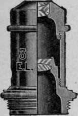
Fig. 10.
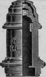
Fig. 11.
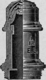
Fig. 12. Objectives.
Fig. 13.
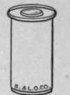
Fig. 14.
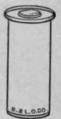
Fig. 15. Eye-Pieces.
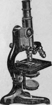
Fig. 16. Pharmacognostic Microscope.
Continue to:
My Books

