Compound Microscopes. Part 2
Description
This section is from the "Histology of Medicinal Plants" book, by William Mansfield. Also see Amazon: Histology of Medicinal Plants.
Compound Microscopes. Part 2
Forms Of Compound Microscopes
The following descriptions refer to three different models of compound microscopes: one which is used chiefly as a pharmacognostic microscope, one as a research microscope stand, while the third type represents a research microscope stand of highest order, which is used at the same time for taking microphotographs.
Pharmacognostic Microscope
The pharmacognostic microscope (Fig. 16) is an instrument which embodies only those parts which are most essential for the examination of powdered drugs, bacteria, and urinary sediments. This microscope is provided with a stage of the dimensions 105 x 105 mm. This factor and the distance of 80 mm. from the optical centre to the handle arm render it available for the examination of even very large objects and preparations, or preparations suspended in glass dishes. The stand is furnished with a side micrometer, a fine adjustment having knobs on both sides, thereby permitting the manipulation of the micrometer screw either by left or right hand. The illuminating apparatus consists of the Abbe condenser of numerical aperture of 1.20, to which is attached an iris diaphragm for the proper adjustment of the light. A worm screw, mounted in connection with the condenser, serves for the raising and lowering of the condenser, so that the cone of illuminating pencils can be arranged in accordance to the objective employed and to the preparation under observation. The objectives necessary are those of the achromatic type, possessing a focal length of 16.2 mm. and 3 mm. Oculars which render the best results in regard to magnification in connection with the two objectives mentioned are the Huyghenian eye-pieces II and IV so that magnifications are obtained varying from 62 to 625. It is advisable, however, to have the microscope equipped with a triple revolving nose-piece for the objectives, so that provision is made for the addition of an oil-immersion objective at any time later should the microscope become available for bacteriological investigations.
The Research Microscope
The research microscope used in research work (Fig. 17) must be equipped more elaborately than the microscope especially designed for the use of the pharmacognosist. While the simple form of microscope is supplied with the small type of Abbe condenser, the research microscope is furnished with a large illuminating apparatus of which the iris diaphragm is mounted on a rack and pinion, allowing displacement obliquely to the optical centre, also to increase resolving power in the objectives when observing those objects which cannot be revealed to the best advantage with central illumination. Another iris is furnished above the condenser; this iris becomes available the instant an object is to be observed without the aid of the condenser, in which case the upper iris diaphragm allows proper adjustment of the light. The mirror, one side plane, the other concave, is mounted on a movable bar, along which it can be slid - another convenience for the adjustment of the light. The microscope stage of this stand is of the round, rotating and centring pattern, which permits a limited motion to the object slide: The rotation of the microscope stage furnishes another convenience in the examination of objects in polarized light, allowing the preparation to be rotated in order to distinguish the polarization properties of the objects under observation.
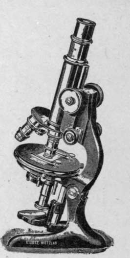
Fig. 17. Research Microscope.
Special Research Microscope
A special research microscope of the highest order (Fig. 18) is supplied with an extra large body tube, which renders it of special advantage for micro-photography. Otherwise in its mechanical equipment it resembles very closely the medium-sized research microscope stand, with the exception that the stand is larger in its design, therefore offering universal application. In regard to the illuminating apparatus, it is advisable to mention that the one in the large research microscope stand is furnished, with a three-lens condenser of a numerical aperture of 1.40, while the medium-sized research stand is provided with a two-lens condenser of a numerical aperture of 1.20. The stage of the microscope is provided with a cross motion - the backward and forward motion of the preparation is secured by rack and pinion, while the side motion is controlled by a micrometric worm screw. In cases where large preparations are to be photographed, the draw-tube with ocular and the slider in which the draw-tubes glide are removed to allow the full aperture of wide-angle objectives to be made use of.
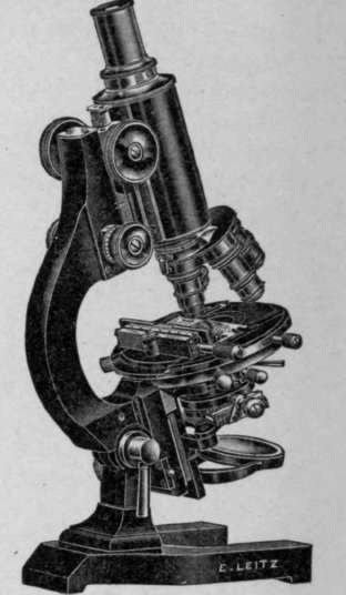
Fig. 18. Special Research Microscope.
Binocular Microscope
The Greenough binocular microscope, as shown in Fig. 19, consists of a microscope stage with two tubes mounted side by side and moving on the same rack and pinion for the focusing adjustment. Either tube can be used without the other. The oculars are capable of more or less separation to suit the eyes of different observers. In each of the drub-like mountings, near the point where the oculars are introduced, porro-prisms have been placed, which erect the image. This microscope gives most perfect stereoscopic images, which are erect instead of inverted, as in the monocular compound microscopes. The Greenough binocular microscope is especially adapted for dissection and for studying objects of considerable thickness.
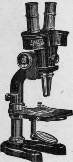
Fig. 19. Greenough Binocular Microscope.
Continue to:
My Books

