Chapter III. Microscopic Measurements
Description
This section is from the "Histology of Medicinal Plants" book, by William Mansfield. Also see Amazon: Histology of Medicinal Plants.
Chapter III. Microscopic Measurements
In making critical examinations of powdered drugs, it is frequently necessary to measure the elements under observation, particularly in the case of starches and crystals.
Ocular Micrometer
Microscopic measurements are made by the ocular micrometer (Fig. 21). This consists of a circular piece of transparent glass on the centre of which is etched a one- or two-millimeter scale divided into one hundred or two hundred divisions respectively. The value of each line is determined by standardizing with a stage micrometer.

Fig. 21. Ocular Micrometer.
Stage Micrometer
The stage micrometer (Fig. 22) consists of a glass slide upon which is etched a millimeter scale divided into one hundred equal parts or lines: each line has a value of one hundredth of a millimeter.
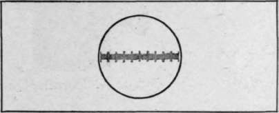
Fig. 22. Stage Micrometer.
Standardization Of Ocular Micrometer With Low-Power. Objective
Having placed the ocular micrometer in the eye-piece and the stage micrometer on the centre of the stage, focus until the lines of the stage micrometer are clearly seen. Then adjust the scales until the lines of the stage micrometer are parallel with and directly under the lines of the ocular micrometer.
Ascertain the number of lines of the stage micrometer covered by the one hundred lines of the ocular micrometer. Then calculate the value of each line of the ocular. This is done in the following manner:
If the one hundred lines of the ocular cover seventy-five lines of the stage micrometer, then the one hundred lines of the ocular micrometer are equivalent to seventy-five one-hundredths, or three-fourths, of a millimeter. One line of the ocular micrometer will therefore be equivalent to one-hundredth of seventy-five one-hundredths, or .0075 part of a millimeter, and as a micron is the unit for measuring microscopic objects, this being equivalent to one one-thousandth of a millimeter, the value of each line of the ocular will therefore be 7.5 microns.
With the high-power objective in place, ascertain the value of each line of the ocular. If one hundred lines of the ocular cover only twelve lines of the stage micrometer, then the one hundred lines of the ocular are equivalent to twelve one-hun-dredths of a millimeter, the value of one line being equivalent to one one-hundredth of twelve one-hundredths, or twelve ten-thousandths of a millimeter, or .0012, or 1.2 u.
It will therefore be seen that objects as small as a thousandth of a millimeter can be accurately measured by the ocular micrometer.
In making microscopic measurements it is only necessary to find how many lines of the ocular scale are covered by the object. The number of lines multiplied by the equivalent of each line will be the size of the object in microns, or micro-millimeters.
Micrometer Eye-Pieces
Micrometer eye-pieces (Figs. 23 and 24) may be used in making measurements. These eye-pieces with micrometer combinations are preferred by some workers, but the ocular micrometer will meet the needs of the average worker.

Fig. 23. Micrometer Eye-Piece.

Fig. 24. Micrometer Eye-Piece.
Mechanical Stages
Moving objects by hand is tiresome and unsatisfactory, first, because of the possibility of losing sight of the object under observation, and secondly, because the field cannot be covered so systematically as when a mechanical stage is used for moving slides.
The mechanical stage (Fig. 25) is fastened to the stage by a screw. The slide is held by two clamps. There is a rack and pinion for moving the slide to left or right, and another rack and pinion for moving the slide forward and backward.
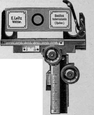
Fig. 25. Mechanical Stage.
Camera Lucida
The camera lucida is an optical mechanical device for aiding the worker in making drawings of microscopic objects. The instrument is particularly necessary in research work where it is desirable to reproduce an object in all its details. In fact, all reproductions illustrating original work should be made by means of the camera lucida or by microphotography.
A great many different types of camera lucidas or drawing apparatus are obtainable, varying from simple-inexpensive to complex-expensive forms. Figs. 26, 27, and 28 show simple and complex forms.

Fig. 26. Camera Lucida.
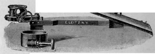
Fig. 27. Camera Lucida.
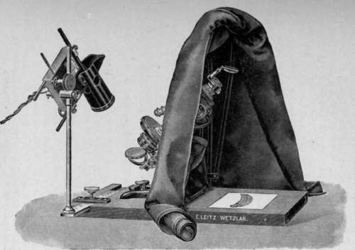
Fig. 28. Drawing Apparatus.
Microphotographic Apparatus
The microphotographic apparatus (Fig. 29), as the name implies, is an apparatus constructed in such a manner that it may be attached to a microscope when we desire to photograph microscopic objects. It consists of a metal base and a polished metal pillar for holding the bellows, slide holder, ground-glass observation plate, and eye-piece. In making photographs, the small end of the bellows is attached to the ocular of the microscope, the focus adjusted, and the object or objects photographed. More uniform results are obtained in making such photographs if an artificial light of an unvarying candle-power is used.

Fig. 29. Microphotographic Apparatus.
There are obtainable more elaborate microphotographic apparatus than the one figured and described, but for most workers this one will prove highly satisfactory. It is possible, by inclining the tube of the microscope, to make good micro-photographs with an ordinary plate camera. This is accomplished by removing the lens of the camera and attaching the bellows to the ocular, focusing, and photographing.
Continue to:
My Books

