Chapter VI. Aerating Tissue
Description
This section is from the "Histology of Medicinal Plants" book, by William Mansfield. Also see Amazon: Histology of Medicinal Plants.
Chapter VI. Aerating Tissue
The aerating tissue of the plant performs a threefold function: first, it permits the exchange of gases during photosynthesis; secondly, it permits the entrance of oxygen and the exit of carbon dioxide during respiration; and, thirdly, it permits the exit of the excess of water absorbed by the plant.
The above functions are carried on by the stomata, the water-pores, the lenticels, and the intercellular spaces of the plant. The stoma functions as the chief channel for the passage of C02-laden air into the leaf and of oxygen-laden air from the leaf to the atmosphere. The stoma also functions as an organ of transpiration, since through the stoma a large part of the excess water of the plant passes off into the air.
Water-Pores
In certain plants the primary epidermis is provided with openings resembling stomata, but unlike stomata the orifice remains open, and instead of being located on the upper or lower surface of the leaf, they are located on the margin of leaves immediately outward from the veins. Water is given off to the atmosphere from these openings. Such an opening is usually designated as a water-pore.
Stomata
The chief external openings of the epidermis of leaves, of herbs, and of young wood stems are known as stomata. Surrounding the stoma are two cells known as guard cells.
Guard cells differ greatly in form, in size, in arrangement, in occurrence, in association, in abundance (Plates 53, 54, and 55), and in color. The guard cells surrounding the stoma vary in form from circular to lens-shaped. In most leaves the outline of the guard cells is rounded or has a curved outline; but in a few cases the guard cells have angled outlines.

Plate 53. 1. Stoma and surrounding cells of aconite stem (Aconitum napellus, L.).
2. Stoma and angled striated walled surrounding cells of peppermint stem (Mentha piperita, L.). 3. Stoma and elongated surrounding cells of lobelia stem (Lobelia inflata, L.).

Plate 54. Types of Stoma.
1. Under epidermis of short buchu (Barosma betulina, [Berg.] Bartlingand Wendl., f.) showing stoma and deposits of hesperidin.
2. Under epidermis of Alexandria senna (Cassia acutifolia, Delile) showing stoma and thick-angled walled surrounding cells.
3. Upper epidermis of eucalyptus leaf (Eucalyptus globulus, Labill.) showing sunken stoma and slightly beaded walled surrounding cells.
4. Under epidermis of belladonna leaf (Atropa belladonna, L.) showing stoma and wavy, striated, walled epidermal cells.
The arrangement of the surrounding cells of the stoma is one of the most important characteristics of the different leaves. As a rule the number of surrounding cells about a stoma is constant for a given species. In senna leaves (Plate 54, Fig. 2) there are normally two surrounding cells about each guard cell, while in coca there are four (Plate 55, Fig. 1). In senna the long diameter of the surrounding cells is parallel to the long diameter of the guard cells; but in coca the long diameter of two surrounding cells is at right angles to the long diameter of the guard cells, while two cells are parallel to the long diameter of the guard cells.
In most leaves there are more than two cells around the guard cells.
The form and size of the surrounding cells must always be considered. In most leaves they are variable in size and form.
Guard cells occur first, even with the surface of the leaf (Plate 56, Fig. A); secondly, above the surface of the leaf (Plate 56, Fig. B); and, thirdly, below the surface of the leaf. (Plate 56, Fig. C). Only one of the above types occurs in a given species of plant. That is, plants with stomata above the surface of the leaf do not have stomata on a level with or below the leaf surface.
The number of stomata on a given surface of a different leaf varies considerably.
In many of the medicinal leaves stomata occur only on the under surface of the leaf. In other leaves stomata occur on both surfaces of the leaf; but in such cases there are a greater number on the under surface.
In certain leaves the long diameter of the guard cells is parallel to the length of the leaf; in other cases the long diameter of the stoma is arranged at right angles to the length of the leaf.
In other leaves the arrangement is still more irregular, the guard cells assuming all sorts of positions in relation to the length of the leaf.
The relation of the stoma to surrounding cells is best shown in cross-sections of the leaf. In powders the relationship of the stoma to the surrounding cells is, however, readily ascertained. If the guard cells come in focus first, they are above the surface; if the guard cells and the surrounding cells come in focus at the same time, the stomata are even with the surface; if the stomata come in focus after the surrounding cells, they are below the surface of the leaf. The relationship of the stoma to the surrounding cells should always be ascertained, not only in cross-sections of the leaf, but also in powders.

Plate 55. Leaf Epidermi with Stoma.
1. Under epidermis of coca leaf (Erythroxylon coca, Lam.) with stoma on a level with the surface.
2. Under epidermis of false buchu (Marrubium peregrinum, L.) with stoma below the level of the surface.
3. Upper epidermis of deer tongue (Trilisia odoratissima, [Wait.] Cass.) with stoma above the leaf surface.
There is the greatest possible variation in the size of guard cells. This fact must always be kept in mind when studying leaves. This variation in the size of the guard cells is clearly illustrated by coca, senna, and by deer's-tongue. In coca the stomata are very small; in senna they are larger; while in deer's-tongue the stomata are very large.
The width and length of the stoma or opening between the guard cells are of a character which must not be overlooked. Generally speaking, those leaves which have large guard cells will have correspondingly large stomata.
The guard cells usually contain chloroplasts showing various stages of decomposition.
In bay-rum leaf the guard cells are of a bright reddish-brown color, but in most leaves the guard cells are colorless.
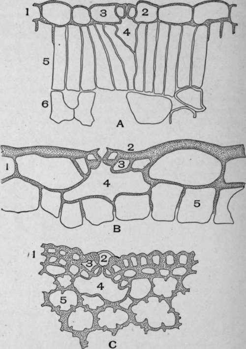
Plate 56. A. Cross-section of belladonna leaf (Atropa belladonna, L.). I, Epidermal cells; 2, Guard cells even with the leaf surface; 3, Surrounding cells; 4, Air space below the guard cells; 5, Palisade cells; 6, Mesophyll cells. B. Cross-section of deer tongue leaf. I. Epidermal cells; 2, Guard cells above the surface of the leaf; 3, Surrounding cells; 4, Air space below the guard cells; 5, Hypodermal cells. C. Cross-section of white pine leaf (Pinus strobus, L.). 1, Epidermal and hypodermal cells; 2, Guard cells below the leaf surface; 3, Surrounding cells; 4, Air space below the guard cells; 5, Parenchyma cells with projecting inner walls.
Continue to:
My Books

