Synthetic Tissue. Continued
Description
This section is from the "Histology of Medicinal Plants" book, by William Mansfield. Also see Amazon: Histology of Medicinal Plants.
Synthetic Tissue. Continued
Synthetic Tissue. Secretion Cavities
Secretion cavities are divided into three groups, according to the nature of the origin of the cavity: first, schizogenous cavities, which originate by a separation of the walls of the secretion cells; secondly, lysigenous cavities, which arise by the dissolution of the walls of centrally located secretion cells; and thirdly, schizo-lysigenous cavities, which originate schizogen-ously, but later become lysigenous owing to the dissolution of the outer layers of the secretion cells.
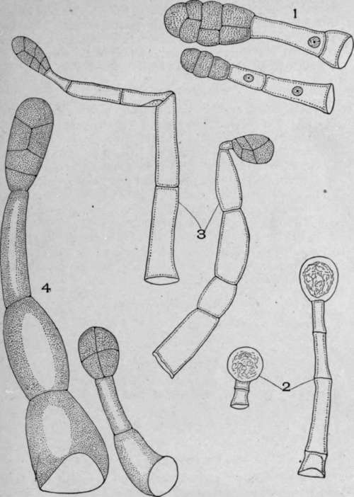
Plate 61. Stalked Glandular Hairs.
1. Belladonna leaf (Atropa belladonna, L.).
2. Geranium stem (Geranium maculatum, L.).
3. Henbane leaf (Hyoscyamus niger, L.).
4. Tobacco leaf (Nicotiana tabacum, L.).
Schizogenous Cavities
Schizogenous cavities occur in white pine bark (Plate 62, Fig. B). The cells lining the cavity are mostly tangentially elongated, and the wall extends into the cavity in the form of a papillate projection. Immediately back from these cells are two or three layers of cells which resemble cortical parenchyma cells, except that they are smaller and their walls are thinner.
' In white pine bark there is a single layer of thin-walled cells lining the cavity. Immediately surrounding the secretion cells is a single layer of thick-walled fibrous cells.
In klip buchu (Plate 63, Fig. B), as in white pine leaf (Plate 64, Fig. B), there is a single layer of thin-walled secretion cells which are surrounded on three sides with parenchyma cells and on the outer side by epidermal cells.
Lysigenous Cavities
Lysigenous cavities occur on the rind of citrus fruits - bitter and sweet orange, lemon, grapefruit, lime, etc., and in the leaves of garden rue, etc.
In bitter orange peel. (Plate 64, Fig. A) the cavity is very large, and the cells bordering the cavity are broken and partially dissolved. The entire cells back of these are white, thin-walled, tangentially elongated cells. There is a great variation in the size of these cavities, the smaller cavities being the recently formed cavities.
Schizo-Lysigenous Cavities
Schizo-lysigenous cavities are formed in white pine bark and many other plants owing to the increase in diameter of the stem. In such cases the walls of the secreting cells break down. The resulting cavity resembles lysigenous cavities.
Unicellular secretion cavities occur in ginger, aloe, calamus, and in canella alba barb.
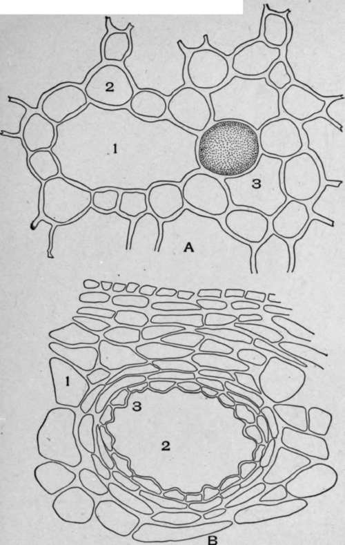
Plate 62. A. Cross-section of calamus rhizome" (Acorus calamus, L.). 1, Intercellular space; 2, Parenchyma cells; 3, Secretion cavity. B. Cross-section of white pine bark (Pinus strobus, L.). 1, Parenchyma; 2, Secretion cavity; 3, Secretion cells.
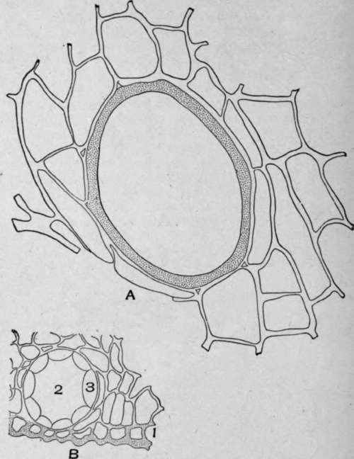
Plate 63. A. Cross-section of a portion of canella alba bark (Canella alba, Murr.).
1. Excretion cavity. B. Cross-section of a portion of klip buchu leaf.
1. Epidermal cells.
2. Secretion cavity.
3. Secretion cells.
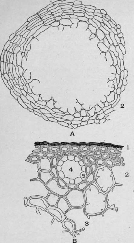
Plate 64. A, Cross-section of bitter orange peel (Citrus aurantium, amara, L.). 1, Internal secretion cavity formed by the dissolution of the walls of the central secreting cells; 2, Secretion cells. B. Cross-section of white pine leaf (Pinus strobus, L.). 1, Epidermal and hypodermal cells; 2, Parenchyma cells with protuding inner walls; 3, Endodermis; 4, Secretion cavity; 5, Secretion cells.
In calamus (Plate 62, Fig. A) the cavity is larger than the surrounding cells; it is rounded in outline, and it contains oleoresin. These cavities are in contact with the ordinary parenchyma cells, from which they are easily distinguished by their larger size and rounded form.
The unicellular oil cavity of canella alba (Plate 63, Fig. A) is rounded or oval in cross-section and is many times larger than the surrounding cells. The wall, which is very thick, is of a yellowish color.
Secretion cavities vary greatly in form, according to the part of the plant in which they are found. In flower petals and leaves they are spherical; in barks they are usually elliptical; in umbelliferous fruits they are elongated and tube-like.
Mucilage cavities are not of common occurrence in medicinal plants. They occur, however, in the stem and root bark of sassafras, the stem bark of slippery elm, the root of althea, etc.
Continue to:
My Books

