X. Effects Of Diet On The Development And Structure Of The Uterus
Description
This section is from the book "Food And Feeding In Health And Disease", by Chalmers Watson. Also available from Amazon: Food and Feeding in Health and Disease.
X. Effects Of Diet On The Development And Structure Of The Uterus
The alarming and persistent decline in the birth-rate is a matter of national interest the importance of which can hardly be overestimated. The purely medical aspect of this question is, however, exceedingly difficult of approach, since social factors and social selfishness have undoubtedly a material influence. During the last generation, however, there has undoubtedly been a marked change in the relative proportions of the constituents of the average man's diet. Can such a change of diet have any influence on the birth-rate?
The following investigation was undertaken to discover what changes, if any, were produced in the development and structure of the uterus by various diets.
The animals employed in the research were rats. The uteri of eighty-six animals were examined, both macroscopically and microscopically. The tissue for microscopic examination was removed in all cases from, as nearly as possible, the same area - the junction of the distal and mid thirds of the uterus.
Eight wild rats, in various stages of development, from the immature to the adult animal, were examined in order to form an opinion as to the structure of the uterus in animals living, presumably, under natural conditions.
The remaining animals were divided into five series, namely: -
(a) A series of thirteen animals fed, from weaning, for periods of from nine to fourteen weeks on an exclusively milk diet. In one section of this series plain milk was used; in another, pasteurised milk; in the third, sterilised milk.
(b) A series of twenty-seven animals fed for periods of from twenty-one days to nine months on bread soaked in milk.
(c) A series of eleven animals fed for periods of from four to fourteen weeks on a rice diet.
(d) A series of five animals fed for varying periods on a diet of porridge or oats.
(e) A series of twenty-two animals fed for periods of from twenty days to eight months on a raw-meat diet.
In the cases of the raw-meat and rice diets, some animals were put on the diet as soon as weaned, others after they had reached various stages of development.
The uterus of the adult wild rat is lined by columnar epithelium. There are glands lined by epithelium which varies from low cubical to columnar in type. The mucous coat is bounded externally by a muscular coat. In the mucous layer three varieties of cells are found: -
1. A cell with a large round or oval, relatively faintly-staining nucleus. This appears to be a young connective tissue type of cell.
2. A cell with a small, round, darkly-staining nucleus, comparable to a lymphoid cell.
1 Malcolm Campbell, F.R.C.S., British Medical Journal, vol. i., 1907
3. A cell with an elongated, very darkly staining nucleus, similar to cells got in fibrous tissue.
In the wild tat the large cells are most numerous. In the wild rat the cells of the other types are few in number, and are chiefly found in the part of the mucosa near the muscular coat.
From the examination of this series of eighty-six animals it is evident that in animals of the same age and approximately of the same weight, living under similar conditions, the uteri may vary within a limited range in size and development. The muscular coat is relatively uniform. The mucosa shows the greatest variations. While in most cases the epithelium lining the cavity is columnar, in some it is cubical; there are also found marked variations as to the position of the nucleus and its staining reactions.
The animals fed on milk and bread soaked in milk approximate most nearly to the type of structure got in the wild rat. In the other groups, fed on what we may term "abnormal diets" - namely, rice, porridge, oats or raw meat - there is found a relatively constant departure from the normal. The type of change is common to all abnormal diets; its severity varies.
The severity of the changes induced are found to be in proportion to the ages of the animals at the time when the abnormal diet was begun. The changes are most marked in the animals put on the diet at weaning; they are less marked the more mature the animal at the time of the commencement of the abnormal diet. In a fully-developed animal, any abnormal diet may fail to materially change either the size or structure of the uterus.
In regard to the development of the uterus, an abnormal diet appears to arrest the growth of the uterus. This arrest of development is most marked in animals fed from weaning on ox-flesh, but is also very well seen in animals fed on rice or on porridge or oats.
In regard to structure, all the abnormal diets led to a diminution of the number of the large connective-tissue type of cells, and a relative increase in the small cells. This change, which may be described as a fibrosis, was most marked in animals fed from weaning on an ox-flesh diet for periods of from four to five months; in this group of animals none became pregnant, while controls from the same litters, fed on bread and milk, all had young.
From these observations it seems justifiable to state: -
1. The use of a non-physiological diet - for example, exclusive flesh, rice, or porridge - induces in the great majority of cases a modification in the structure of the uterine mucous membrane. This modification consists in a diminution in the number of the large connective tissue type of cells, which appear to be important constituents in a physiologically active mucosa.
2. The structural change is most profound in animals fed from weaning on an exclusively ox-flesh diet. In such animals the development of the uterus is also most interfered with.
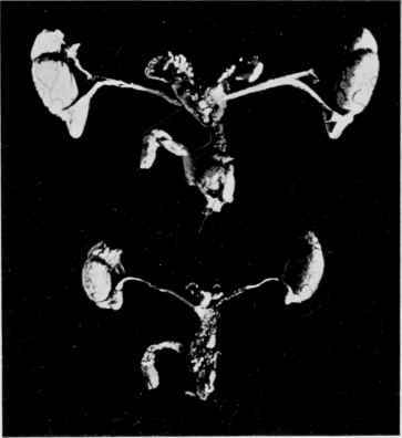
Fig. 10. - Male Reproductive Organs of full-grown Rats from the same litter.
A. After 5 1/2 months, bread-and-milk diet. Weight of animal 210 grms. B. After 5 1/2 months, ox-flesh. Weight of animal 170 grms.
(Face page 588.
3. The structural change in two is associated with sterility. Chalmers Watson, to whom I am indebted for much of the material used in this investigation, pointed out that a meat diet, if begun at weaning, almost invariably led to sterility. The present investigation shows that the sterility is probably due to the structural and developmental abnormalities in the uterus induced by the abnormal diet. It has been shown that the consumption of meat per head in this country is to-day almost seventeen times as great as it was in 1850. During the same period the fall in the birth-rate has been most marked.
While it would be unwarrantable to attempt to found any theories in regard to the falling birth-rate on the results of this limited experiment, yet, when we add to the above facts the further consideration that the diminished birth-rate is most marked in the better-off classes of society - that is, among the very classes whose means permit of an unrestricted use of the more expensive meat diet - it is evident that the food factor is one which is at least worthy of consideration in dealing with the great question of the falling birth-rate.
Continue to:
My Books

