XVII. The Changes In The Structure Of The Thyroid Gland IX Wild Rats Under The Influence Of Altered Dietetic Conditions
Description
This section is from the book "Food And Feeding In Health And Disease", by Chalmers Watson. Also available from Amazon: Food and Feeding in Health and Disease.
XVII. The Changes In The Structure Of The Thyroid Gland IX Wild Rats Under The Influence Of Altered Dietetic Conditions
In former papers the author has shown that diet modifies the minute structure of the thyroid gland in tame rats. The following investigation was undertaken to ascertain to what extent, if any, the thyroid of wild rats is influenced by a diet which is unusual for these animals. A series of twenty young wild rats, apparently healthy, was obtained from a hotel basement. (For reasons given later it is essential in such an experiment that all the rats be obtained from the same source and preferably at the same time.) Ten of the series, of weights ranging from 60 to 130 grammes, were killed on receipt; the remaining ten were fed in the laboratory for ten weeks on a diet of bread soaked in skimmed milk.2 Under this regime the animals seemed to maintain perfect health, although it was observed that for a time their growth was retarded; their weight, when killed, ranged from no to 170 grammes. The thyroid glands were fixed in 5 per cent, formalin; the glands were weighed on the second day, the excess of fluid having previously been removed; one gland from each rat was carried through in the usual manner and stained with haematoxylin and eosin, a section of each series being stained on the same slide. The average percentage weight of the gland of the rats in Series 1 - those killed on receipt - was .0202 gramme, that of Series 2 - after ten weeks' feeding in the laboratory - .0265 gramme, which represents an appreciable increase in weight. There is a striking contrast in the histological appearances of the gland in the two series. In Series 1, where the appearances are uniform throughout, the vesicles are large and full of colloid which takes on a delicate pink stain with the haematoxylin and eosin stain; the secreting cells lining the vesicles are flat in character, and consist of a small darkly stained nucleus with a small amount of protoplasm in the body of the cell (Fig 29). In Series 2, where the picture is also uniform, the histological appearances are different from the above. The vesicles are for the most part small and contain little or no secretion. If colloid is present it appears as a thin unstained secretion, or as a faintly stained granular colloid. The cells lining the vesicles are large and situated at the basal part of the cell, there being a free margin of faintly granular protoplasm. The blood-vessels are more prominent throughout (Fig. 30). It should be added that in two of this series the histological appearances are intermediate in character between those represented in Figs. 1 and 2. The striking difilerences in the histological appearances of the glands in the two series might be due to either (a) altered environment or (b) the changed diet. The former explanation seems not to hold good, since I have observed that in some wild rats obtained from a different source, and which were killed on receipt, the histological appearances of the thyroid gland were identical with those described in Series 2. The second explanation is therefore the more probable, and we have, I believe, in these results evidence of a modification in structure and function of the thyroid gland induced by diet.
1 Chal mers Watson, Journal of Physiology, vol. xxxv., 1907. 2 It is essential that the milk be not given in excess.
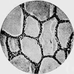
Fig. 29. - Thyroid Gland of Wild Rat (caught in a hotel basement) which was killed on receipt.
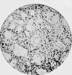
Fig. 30. - Thyroid Gland of Wild Rat from same source as Fig. 29 after feeding for ten weeks on a bread-and-milk diet.
[Face page 604.
Continue to:
My Books

